

CHEMICAL PROPERTIES OF MONOSACCHARIDES. STRUCTURE, COMPOSITION AND PROPERTIES OF DISACCHARIDES. STRUCTURE, COMPOSITION AND PROPERTIES OF POLYSACCHARIDES.
Introduction: Carbohydrates are the most abundant class of bioorganic molecules on planet Earth. Although their abundance in the human body is relatively low, carbohydrates constitute about 75% by mass of dry plant materials.
Green (chlorophyll-containing) plants produce carbohydrates via photosynthesis. In this process, carbon dioxide from the air and water from the soil are the reactants, and sunlight absorbed by chlorophyll is the energy source.
Carbohydrates are the most abundant class of organic compounds found in living organisms. They originate as products of photosynthesis, an endothermic reductive condensation of carbon dioxide requiring light energy and the pigment chlorophyll

|
n
CO2 + n H2O + energy |
As noted here, the formulas of many carbohydrates can be written as carbon hydrates, Cn(H2O)n, hence their name. The carbohydrates are a major source of metabolic energy, both for plants and for animals that depend on plants for food. Aside from the sugars and starches that meet this vital nutritional role, carbohydrates also serve as a structural material (cellulose), a component of the energy transport compound ATP, recognition sites on cell surfaces, and one of three essential components of DNA and RNA.
Plants have two main uses for the carbohydrates they produce. In the form of cellulose, carbohydrates serve as structural elements, and in the form of starch, they provide energy reserves for the plants.
Dietary intake of plant materials is the major carbohydrate source for humans and animals. The average human diet should ideally be about two-thirds carbohydrate by mass.
Carbohydrates have the following functions in humans:
1. Carbohydrate oxidation provides energy.
2. Carbohydrate storage, in the form of glycogen, provides а short- term energy reserve.
3. Carbohydrates supply carbon atoms for the synthesis of other biochemical substances (proteins, lipids, and nucleic acids).
4. Carbohydrates form part of the structural framework of DNA and RNA molecules.
5. Carbohydrate "markers" on cell surfaces play key roles in cell -cell recognition processes.
CARBOHYDRATE CLASSIFICATIONS
Most simple carbohydrates have empirical formulas that fit the general formula СnН2nОn. An early observation by scientists that this general formula can also be written as Сn(Н2О)n is the basis for the term carbohydrate - that is, "hydrate of carbon." It is now known that this hydrate viewpoint is not correct, but the term carbohydrate still persists. Today the term is used to refer to an entire family of compounds, only some of which have the formula СnН2nОn.
Carbohydrates are polyhydroxy aldehydes, polyhydroxy ketones, or compounds that yield such substances upon hydrolysis. The carbohydrate glucose is а polyhydroxy aldehyde, and the carbohydrate fructose is а polyhydroxy ketone.
А striking structural feature of carbohydrates is the large number of functional groups present. In glucose and fructose there is а functional group attached to each carbon atom. Carbohydrates are classified on the basis of molecular size as monosaccharides, oligosaccharides, and polysaccharides.
Monosaccharides are carbohydrates that contain a single polyhydroxy aldehyde or polyhydroxy ketone unit. Monosaccharides cannot be broken down into simpler units by hydrolysis reactions. Both glucose and fructose are monosaccharides. Naturally occurring monosaccharides have4rom three to seven carbon atoms; five- and six-carbon species are especially common. Pure monosaccharides are water-soluble, white, crystalline solids.
Oligosaccharides are carbohydrates that contain from two to ten monosaccharide units. Disaccharides are the most common type of oligosaccharide. Disaccharides are carbohydrates composed of two monosaccharide units covalently bonded to each other. Like monosaccharides, disaccharides are crystalline, water-soluble substances. Sucrose (table sugar) and lactose (milk sugar) are disaccharides.
Within the human body, oligosaccharides are often found associated with proteins and lipids in complexes that have both structural and regulatory functions. Free oligosaccharides, other than disaccharides, are seldom encountered in biological systems.
Complete hydrolysis of an oligosaccharide produces monosaccharides. Upon hydrolysis, а disaccharide produces two monosaccharides, а trisaccharide three monosaccharides, а hexasaccharide six monosaccharides, and so on.
Polysaccharides are carbohydrates made up of many monosaccharide units. Polysaccharides, which are polymers, often consist of tens of thousands of monosaccharide units. Both cellulose and starch are polysaccharides. We encounter these two substances everywhere. The paper on which this book is printed is mainly cellulose, as are the cotton in our clothes and the wood in our houses. Starch is а component of many types of foods including bread, pasta, potatoes, rice, corn, beans, and peas.
Chirality: handedness in molecules
Before considering structures for and reactions of specific carbohydrates, we will consider handedness, а biologically important structural property exhibited by most carbohydrates. Most carbohydrate molecules can exist in two forms - а left-handed form and а right-handed form. Significantly, these different forms often elicit different responses within the human body.
Mirror Images. The concept of mirror images is the key to understanding molecular handedness. All objects, including all molecules, have mirror images. The mirror image of an object is the object’ reflection in а mirror. For example: human hands.
Chirality. Objects that cannot be superimposed upon their mirror image are said to be chiral objects. А chiral object is an object that is not identical to its mirror image. Your hands and feet are chiral objects, as are gloves and shoes. Objects that can be superimposed upon their mirror images are achiral. An achiral object is identical to its mirror image. Achiral objects include tube socks, solid-colored ties and Т-shirts.
Molecules, like larger objects, can be chiral or achiral. А simple example of а chiral molecule is the trisubstituted methane bromochloroiodomethane.
The simplest example of а chiral carbohydrate is the three-carbon molecule glyceraldehyde.
Trying to superimpose the mirror image of а molecule on that molecule visually, is one way to determine molecular chirality. Another method, which is much easier to apply, makes use of the observation that generally, whenever а carbon atom in а molecule is bonded to four different groups, the molecule as а whole is chiral.
Any organic molecule containing а single carbon atom with four different groups attached to it exhibits chirality. Such а carbon atom is called а chiral center. А chiral center is an atom in а molecule that has four different groups tetrahedrally bonded to it.
Chiral centers within molecules are often denoted by а small asterisk. Note the chiral centers in the following molecules.
2-butanol 1-chloto-1-iodoethane 3-methylhexane
Organic molecules, especially carbohydrates, may contain more than one chiral center. For example, the following carbohydrate has two chiral centers.
Stereoisomers are isomers whose atoms are connected in the same way but differ in their arrangement in space. The two nonsuperimposable mirror-image forms of а chiral molecule are stereoisomers.
There are two major causes of stereoisomerism: (1) the presence of а chiral center in а molecule, and (2) the presence of "structural rigidity" in а molecule. Structural rigidity is caused by restricted rotation about chemical bonds. It is the basis for cis - trans stereoisomerism, а phenomenon found in some substituted cycloalkanes and some alkenes.
Stereoisomers can be subdivided into two types: enantiomers and diastereomers. Enantiomers are stereoisomers whose molecules are nonsuperimposable mirror images of each other. Left- and right-handed forms of а molecule with а single chiral center are enantiomers.
Diastereomers are stereoisomers whose molecules are not mirror images of each other. Cis - trans isomers (of both the alkene and the cycloalkane types) are diastereomers. We will see additional examples of carbohydrate diastereomers in the next section. Stereoisomers that are not enantiomers are diastereomers; they must be one or the other.
Enantiomers Diastereomers
Fischer Projections. Drawing three-dimensional representations of chiral molecules, can be both time-consuming and awkward. Fischer projections represent а method for giving molecular chirality specifications in two dimensions. А Fischer projection is а two-dimensional notation showing the spatial arrangement of groups about chiral centers in molecules.
In а Fischer projection, а chiral center is represented as the intersection of vertical and horizontal lines. The atom at the chiral center, which is almost always carbon, is not explicitly shown.
The tetrahedral arrangement of the four groups attached to the atom at the chiral center is governed by the following conventions: (1) Vertical lines from the chiral center represent bonds to groups directed into the printed page. (2) Horizontal lines from the chiral center represent bonds to groups directed out of the printed page.
Fischer projection
Our immediate concern is Fischer projections for monosaccharides. Such projections have the monosaccharide carbon chain positioned vertically with the carbonyl group (aldehyde or ketone) at or near the top.
The smallest monosaccharide that has а chiral center is the compound glyceraldehydes (2,3-dihydroxypropanal). The structural formula and Fischer projections for the two enantiomers of glyceraldehyde are
D-glyceraldehyde L-glyceraldehyde
The handedness (right and left) of these two enantiomers is specified by using the designations D and L. The enantiomer with the chiral center - ОН group on the right in the Fischer projection is by definition the right-handed isomer (в-glyceraldehyde), and the enantiomer with the chiral center - ОН group on the left in the Fischer projection is by definition the left-handed isomer (L-glyceraldehyde).
We now consider Fischer projections for the compound 2,3,4-trihydroxybutanal, а monosaccharide with four carbons and two chiral centers.
There are four stereoisomers for this compound - two pairs of enantiomers.
A B C D
First enantiomeric pair Second enantiomeric pair
In the first enantiomeric pair, both chiral center - ОН groups are on the same side of the Fischer projection, and in the second enantiomeric pair, the chiral center - ОН groups are on opposite sides of the Fischer projection. These are the only - ОН group arrangements possible.
The D, L system used to designate the handedness of glyceraldehyde enantiomers is extended to monosaccharides with more than one chiral center in the following manner.
A B C D
D-isomer L-isomer D-isomer L- isomer
The carbon chain is numbered, starting at the carbonyl group end of the molecule, and the highest-numbered chiral center is used to determine D or L configuration.
The D, L nomenclature gives the configuration (handedness) only at the highest-numbered chiral center. The configuration at other chiral centers in а molecule is accounted for by assigning а different common name to each pair of D, L enantiomers. In our present example, compounds А and В (the first enantiomeric pair) are D-erythrose and L-erythrose; compounds С and D (the second enantiomeric pair) are D-threose and L-threose.
А and С are diastereomers, stereoisomers that are not mirror images of each other. Other diastereomeric pairs in our example are А and D, В and С, and В and D. These four pairs are epimers. Epimers are diastereomers that differ only in the configuration at one chiral center.
In general, а compound that has n chiral centers may exist in а maximum of 2n stereoisomeric forms. For example, when three chiral centers are present, at most eight stereoisomers (23 = 8) are possible (four pairs of enantiomers).
Stereoisomers. 1. Isomers in which the atoms have the same connectivity but differ in spatial arrangement.
2. Stereoisomerism results either from the presence of а chrial center or from structural rigidity caused by restricted rotation about chemical bonds.
Enantiomers. 1. Stereoisomers that are nonsuperimposable mirror images of each other.
2.Handedness (D and L configuration) is determined by the configuration at the highest-numbered chiral center.
3. Enantiomers rotate plane-polarized light in different directions. (+) Enantiomers are dextrorotatory (clockwise), and (-) enantiomers are levorotatory (counterclockwise).
Diastereomers. 1. Stereoisomers that are not mirror images of each other.
2. Epimers are diastereomers whose configurations differ only at one chiral center.
Properties of Enantiomers. Structural isomers differ in most chemical and physical properties. For example, structural isomers have different boiling points and melting points. Diastereomers also differ in most chemical and physical properties. They also have different boiling points and freezing points. In contrast, nearly all the properties of а pair of enantiomers are the same; for example, they have identical boiling points and freezing points. Enantiomers exhibit property differences in only two areas: their interaction with plane-polarized light and their interaction with other chiral substances.
• An enantiomer that rotates plane-polarized light to the right is said to be dextrorotatory (the Latin dexter means "right"). An enantiomer that rotates plane-polarized light to the left is said to be levorotatory (the Latin laevus means "left").
• А plus or minus sign inside parentheses is used to denote the direction of rotation of plane-polarized light by а chiral compound. The notation (+) means rotation to the right (clockwise), and (-) means rotation to the left (counterclockwise). Thus the dextrorotstory enantiomer of glucose is (+)-glucose.
• An equimolar mixture of two enantiomers is called а racemic mixture, or а racemate. Since а racemic mixture contains equal numbers of dextrorotating and levorotating molecules, the net optical rotation is zero. А racemic mixture is often specified by prefixing the name of the compound with the symbol (± )
Interactions between chiral compounds. А left-handed baseball player (chiral) and а right-handed baseball player (chiral) can use the same baseball bat (achiral) or wear the same baseball hat (achiral). However, left- and right-handed baseball players (chiral) cannot use the same baseball glove (chiral). This nonchemical example illustrates that the chirality of an object becomes important when the object interacts with another chiral object.
Applying this generalization to molecules, we find that the two members of an enantiomeric pair, because of their differing chirality, interact differently with other chiral molecules. We find that:
1. А pair of enantiomers has the same solubility in an achiral solvent, such as ethanol, but differing solubilities in а chiral solvent, such as в-2-butanol.
2. The rate and extent of reaction of enantiomers with another reactant are the same if the reactant is achiral but differ if the reactant is chiral. The different reactions that different enantiomers undergo are further considered in the paragraph that follows.
3. Enantiomers have identical boiling points, freezing points, and densities, because such properties depend on the strength of intermolecular forces, and intermolecular force strength does not depend on chirality. Intermolecular force strength is the same for both forms of а chiral molecule, because both forms have identical sets of functional groups.
The two enantiomeric forms of а chiral molecule often generate different responses from the human body. Sometimes both enantiomers are biologically active, each form giving a different response; sometimes both give the same response, but one isomer’s response is many times greater than that of the other; and sometimes only one of the two forms is biologically active, the other form giving no response. For example, the body’s response to the D isomer of the hormone epinephrine is 20 times greater than its response to the L isomer. Epinephrine exerts its effect by binding to specialized receptors. It binds to the receptor site by means of а three-point contact, D-epinephrine makes а perfect three-point contact with the receptor surface, but the biologically weaker L-epinephrine can make only а two-point contact. Because of the poorer fit, the binding of the isomer is weaker, and less physiological response is observed.
Classification of Monosaccharides. Now that we have considered molecular chirality and its consequences, we return to the subject of carbohydrates by considering further details about monosaccharides, the simplest carbohydrates.
Although there is no limit to the number of carbon atoms that can be present in а monosaccharide, only monosaccharides with three to seven carbon atoms are commonly found in nature. А three-carbon monosaccharide is called а triose, and those that contain four, five, and six carbon atoms are called tetroses, pentoses, and hexoses, respectively.
Monosaccharides are classified as aldoses or ketoses on the basis of type of carbonyl group present. Aldoses are monosaccharides that contain an aldehyde group. Ketoses are monosaccharides that contain а ketone group.
Monosaccharides are often classified by both their number of carbon atoms and their functional group. А six-carbon monosaccharide with an aldehyde functional group is an aldohexose; а five-carbon monosaccharide with а ketone functional group is а ketopentose.
Monosaccharides are also often called sugars. Hexoses are six-carbon sugars, pentoses five-carbon sugars, and so on. The word sugar is associated with "sweetness," and most (but not all) monosaccharides have а sweet taste. The designation sugar is also applied to disaccharides, many of which also have а sweet taste. Thus sugar is а general designation for either а monosaccharide or а disaccharide.
The simplest aldose and ketose are the trioses glyceraldehyde and dihydroxyacetone.
glyceraldehydes dihydroxycaetone
The D and L designations specify the configuration at the highest-numbered chiral center in а monosaccharide. The configurations about other chiral centers are accounted for by assigning а different common name to each pair of D and L enantiomers. This naming system, for simple aldoses, is given
The L forms are mirror images of the molecules shown.
А major difference between glyceraldehyde and dihydroxyacetone is that the latter does not possess а chiral carbon atom. Thus, D and L forms are not possible for dihydroxy acetone. This reduces by half (compared with aldoses) the number of stereoisomers possible for ketotetroses, ketopentoses, and ketohexoses. An aldohexose has four chiral carbon atoms, but а ketohexose has only three. atoins.
Cyclic forms of monosaccharides. So far in this chapter, the structures of monosaccharides have been depicted as open-chain polyhydroxy aldehydes or ketones. However, experimental evidence indicates that for monosaccharides containing five or more carbon atoms, such open-chain structures are actually in equilibrium with two cyclic structures, and the cyclic structures are the dominant forms at equilibrium.
The cyclic forms of monosaccharides result from the ability of their carbonyl group to react intramolecularly with а hydroxyl group. The result is а cyclic hemiacetal or cyclic hemiketal. Such an intramolecular cyclization reaction for D-glucose is shown:
Structure 2 is а rearrangement of the projection formula for D-glucose in which the carbon atoms have locations similar to those found for carbon atoms in а six-membered ring. All hydroxyl groups drawn to the right in the original projection formula appear below the ring. Those to the left in the projection formula appear above the ring.
Structure 3 is obtained by rotating the groups attached to carbon-5 in а counterclockwise direction so that they are in the positions where it is easiest to visualize intramolecular hemiacetal formation. The intramolecular reaction occurs between the hydroxyl group on carbon-5 and the carbonyl group (carbon-1). The - ОН group adds across the carbon - oxygen double bond, producing а heterocyclic ring that contains five carbon atoms and one oxygen atom.
Addition across the carbon - oxygen double bond with its accompanying ring formation produces а chiral center at carbon-l, so two stereoisomers are possible. These two forms differ in the orientation of the - ОН group on the hemiacetal carbon atom (carbon-1). In a-D-glucose, the - ОН group is on the opposite side of the ring from the CH2OH group attached to carbon-5. In b-D-glucose, the СН2ОН group on carbon-5 and the - ОН group on carbon-1 are on the same side of the ring.
In an aqueous solution of в-glucose, а dynamic equilibrium exists among the a, b, and open-chain forms, and there is continual interconversion among them. For example, a freshly mixed solution of pure a-D-glucose slowly converts to а mixture of both a- and b-D-glucose by an opening and а closing of the cyclic structure. When equilibrium is established, 63 % of the molecules are b-D-glucose, 37 % are a-D-glucose, and less than 0.01 % are in the open-chain form.
Intramolecular cyclic hemiacetal formation and the equilibrium between forms associated with it is not restricted to glucose. All aldoses with five or more carbon atoms establish similar equilibria, but with different percentages of the alpha, beta, and open-chain forms. Fructose and other ketoses with а sufficient number of carbon atoms also cyclize; here, cyclic hemiketal formation occurs.
Galactose, like glucose, forms а six-membered ring, but both D-fructose and D-ribose form а five-membered ring.
D-fructose D-ribose
D-Fructose cyclization involves carbon-2 (the keto group) and carbon-5, which results in two CH2OH groups being outside the ring (carbons 1 and 6). D-Ribose cyclization involves carbon-1 (the aldehyde group) and carbon-4.
In а
The D or L form of а monosaccharide is determined by the position of the terminal СН2ОН group on the highest-numbered ring carbon atom. In the в form, this group is positioned above the ring. In the ь form, which is not usually encountered in biological systems, the terminal CH2OH group is positioned below the ring.
a or b configuration is determined by the position of the -ОН group on carbon-1 relative to the CH2OH group that determines D or L series. In а b configuration, both of these groups point in the same direction; in an a configuration, the two groups point in opposite directions.
b-D-Monosaccharide a-D-Monosaccharide b-L-Monosaccharide
Where a or b configuration does not matter, the -ОН group on carbon-1 is placed in a horizontal position, and а wavy line is used as the bond that connects it to the ring.
The specific
identity of а monosaccharide is determined by the positioning of the other: -
ОН groups in the
b-form a-form
Reactions of monosaccharides. Five important reactions of monosaccharides are oxidation, reduction, glycoside formation, phosphate ester formation, and amino sugar formation. In considering these reactions, we will use glucose as the monosaccharide reactant. Remember, however, that other aldoses as well as ketoses undergo similar reactions.
Oxidation. Monosaccharide oxidation can yield three different types of oxidation products. The oxidizing agent used determines the product.
Weak oxidizing agents, such as Tollens, Fehling's, and Benedict's solutions, oxidize the carbonyl group end of а monosaccharide to give an -onic acid. Oxidation of the aldehyde end of glucose produces gluconic acid, and oxidation of the aldehyde end of galactose produces galactonic acid. The structures involved in the glucose reaction are
Because monosaccharides act as reducing agents in such reactions, they are called reducing sugars. With Tollens solution, glucose reduces Ag+ ion to Ag, and with Benedict's and Fehling's solutions, glucose reduces Cu2+ ion to Cu+ ion. А reducing sugar is a carbohydrate that gives a positive test with Tollens, Benedict's and Fehling's solutions. All monosaccharides are reducing sugars.
Tollens, Fehling's, and Benedict's solutions can be used to test for glucose in urine, а symptom of diabetes. For example, using Benedict's solution, we observe that if no glucose is present in the urine (а normal condition), the Benedict's solution remains blue.
The presence of glucose is indicated by the formation of а red precipitate. Testing for the presence of glucose in urine is such а common laboratory procedure that much effort has been put into the development of easy-to-use test methods.
Strong oxidizing agents can oxidize both ends of а monosaccharide at the same time (the carbonyl group and the terminal primary alcohol group) to produce а dicarboxylic acid. Such polyhydroxy dicarboxylic acids are known as -aric acids. For glucose, such an oxidation produces glucaric acid.
Although it is difficult to do in the laboratory, in biological systems enzymes can oxidize the primary alcohol end of an aldose such as glucose, without oxidation of the aldehyde group, to produce а -uronic acid. For glucose, such an oxidation produces D-glucuronic acid.
D-Glucose D-Glucuronic
acid
Reduction. The carbonyl group present in а monosaccharide (either an aldose or а ketose) can be reduced to а hydroxyl group, using hydrogen as the reducing agent. For aldoses and ketoses, the product of the reduction is the corresponding polyhydroxy alcohol, which is sometimes called а sugar alcohol. For example, the reduction D-glucose gives D-glucitol.
D-Glucitol is also known by the common name D-sorbitol. Hexahydroxy alcohols such as D-sorbitol have properties similar to those of the trihydroxy alcohol glycerol. These alcohols are used as moisturizing agents in foods and cosmetics because of their affinity for water. D-Sorbitol is also used as а sweetening agent in chewing gum; bacteria that cause tooth decay cannot use polyalcohols as food sources, as they can glucose and many other monosaccharides.
Mutarotation. a- and b-forms of monosaccharides are readily interconverted when dissolved in water. This spontaneous process, called mutarotation, results in an equilibrium mixture of a- and b-furanose forms and a- and b-pyranose forms. The open chain that is formed during muterotation can participate in oxidation-reduction reactions.
O-Glycoside Formation. As you know, that hemiacetals and hemiketals can react with alcohols in acid solution to produce acetals and ketals. Because the cyclic forms of monosaccharides are hemiacetals and hemiketals, they react with alcohols to form acetals and ketals, as is illustrated for the reaction of b-D-glucose with methyl alcohol.
b-D-glucose Methyl-b-D-glucoside and Methyl-a-D-glucoside
The general name for monosaccharide acetals and ketals is glycoside. А glycoside is an acetal or а ketal forpined pот а cyclic monosaccharide. More specifically, а glycoside produced from glucose is а glucoside, from galactose а galactoside, and so on. Glycosides, like the hemiacetals and hemiketals from which they are formed, can exist in both a and b forms. Glycosides are named by listing the alkyl or aryl group attached to the oxygen, followed by the name of the monosaccharide involved, with the suffix –ide appended to it.
Methyl-a-D-glucoside Methyl-b-D-glucoside
N-Glycoside.
Phosphate ester formation. The hydroxyl groups of а monosaccharide can react with inorganic oxyacids to form inorganic esters. Phosphate esters, formed from phosphoric acid and various monosaccharides, are commonly encountered in biological systems. For example, specific enzymes in the human body catalyze the esterification of the carbonyl group (carbon-1) and the primary alcohol group (carbon-6) in glucose to produce the compounds glucose 1-phosphate and glucose б-phosphate, respectively.
a-D-Glucose-1-phosphate a-D-Glucose-6-phosphate
These phosphate esters of glucose are stable in aqueous solution and play important roles in the metabolism of carbohydrates.
Acylation of Monosaccharides:
Alkylation of Monosaccharides:
Amino Sugar Formation. Amino sugars of glucose, mannose, and galactose are common in nature. Such sugars are produced by replacing the hydroxyl group on carbon-2 on the monosaccharide with an amino group. Amino sugars and their N-acetyl derivatives are important building blocks of polysaccharides found in cartilage.
a-D-Glucosamine a-D-Glalactosamine N-acety1a-D-glucosanune
The N-acetyl derivatives of D-glucosamine and D-galactosamine are present in the biochemical markers on red blood cells, which distinguish the various blood types.
Isomerization. Monosaccharides undergo several types of isomerization, for example, after several hours an alkaline solution of D-glucose will also contain D- mannose and D- fructose. Both isomerizations involve an intramolecular shift of a hydrogen atom and a charge in the location of a double bond.
The intermediate that is formed during this process is called an enediol. The reversible transformation of glucose to fructose is an example of an aldose-ketose interconversion. Because there is a change in the conversion of glucose to mannose is referred to as an epimerization. Several enzyme-catalyzed reactions involving enediols occur in carbohydrate metabolism.
Biologically important monosaccharides. Of the many monosaccharides, the most important in the human body are the D-forms of glucose, galactose, fructose, and ribose. Glucose and galactose are aldohexoses, fructose is а ketohexose, and ribose is an aldopentose. All four of these monosaccharides are water-soluble, white, crystalline solids.
D-Glucose. Of all monosaccharides, о-glucose is the most abundant in nature and the most important from а nutritional standpoint. Its Fischer projection is
D-Glucose
Ripe fruits, particularly ripe grapes (20% - 30% glucose by mass), are а good source of glucose, which is often referred to as grape sugar. Two other names for о-glucose are dextrose and blood sugar. The name dextrose draws attention to the fact that the optically active D-glucose, in aqueous solution, rotates plane-polarized light to the right. The term blood sugar draws attention to the fact that blood contains dissolved glucose. The concentration of glucose in human blood is fairly constant; it is in the range of 70 - 100 mg per 100 mL of blood. Cells use this glucose as а primary energy source.
А 5% (m/v) glucose solution is often used in hospitals as an intravenous source of nourishment for patients who cannot take food by mouth. The body can use it as an energy source without digesting it.
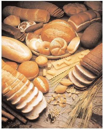
Foods high in carbohydrate include fruits, sweets, soft drinks, breads, pastas, beans, potatoes, bran, rice, and cereals. Carbohydrates are a common source of energy in living organisms; however, no carbohydrate is an essential nutrient in humans.
Carbohydrates are not necessary building blocks of other molecules, and the body can obtain all its energy from protein and fats.[ The brain and neurons generally cannot burn fat for energy, but use glucose or ketones. Humans can synthesize some glucose (in a set of processes known as gluconeogenesis) from specific amino acids, from the glycerol backbone in triglycerides and in some cases from fatty acids. Carbohydrate and protein contain 4 calories per gram, while fats contain 9 calories per gram. In the case of protein, this is somewhat misleading as only some amino acids are usable for fuel.
Organisms typically cannot metabolize all types of carbohydrate to yield energy. Glucose is a nearly universal and accessible source of calories. Many organisms also have the ability to metabolize other monosaccharides and Disaccharides, though glucose is preferred. In Escherichia coli, for example, the lac operon will express enzymes for the digestion of lactose when it is present, but if both lactose and glucose are present the lac operon is repressed, resulting in the glucose being used first. Polysaccharides are also common sources of energy. Many organisms can easily break down starches into glucose, however, most organisms cannot metabolize cellulose or other polysaccharides like chitin and arabinoxylans. These carbohydrates types can be metabolized by some bacteria and protists. Ruminants and termites, for example, use microorganisms to processcellulose
Even though these complex carbohydrates are not very digestible, they represent an important dietary element for humans, called dietary fiber. Fiber enhances digestion, among other benefits.
The biological significance of carbohydrates in living organisms
Two common Monosaccharides, (single sugars) Glucose and Fructose
Sugars are most often found in the form of a "RING". The glucose molecule in the image above and the one in the image below (Glc) are really the same molecule, just arranged differently. The corners of the "stop sign" represent Carbon atoms even thought they are not labeled with a "C" (its chemistry shorthand). To form these rings, the Carbonyl (C=0) Carbon of the straight-chain form (above) forms a bond with the next to last Carbon in the chain, making the ring.
Functions of Carbohydrate
Carbohydrate functions as Bio Fuel
· Polysaccharides such as starch and glycogen are first hydrolyzed by enzymes to Glucose.
· Glucose is then oxidized to produce carbon dioxide and water.
· Energy is released in this process which is used for functioning of the cells.
Carbohydrate functions as Primary Source of Energy
Carbohydrate functions as storage food
Different forms of Carbohydrate are stored in living organism as storage food.
· Polysaccharide starch acts as storage food for plants.
· Glycogen stored in liver and muscles acts as storage food for animals.
· Inulin acts as storage food of dahlias, onion and garlic.
Thus carbohydrate performs the function of storing food.
Carbohydrate functions as framework in body
Different Carbohydrates especially Polysaccharides act as framework in living organism.
· Cellulose forms cell wall of plant cell along with hemicelluloses and Pectin
· Chitin forms cell wall of fungal cell and exoskeleton of arthropods
· Peptidoglycan forms cell wall of bacteria and cyanobacteria.
Thus carbohydrates function as contributing material to the cellular structure.
Carbohydrate functions as Anticoagulant
Carbohydrate functions as Antigen
Carbohydrate functions as Hormone
Carbohydrates provide raw material for industry
D-Galactose. А comparison of the Fischer projections for D-galactose and D-glucose shows that these two compounds differ only in the configuration of the - ОН group and - Н group on carbon-4.]
D-Galactose D-Glucose
D-Galactose and D-glucose are epimers.
D-Galactose is seldom encountered as а free monosaccharide. It is, however, а component of numerous important biochemical substances. In the human body, galactose is synthesized from glucose in the mammary glands for use in lactose (milk sugar), а disaccharide consisting of а glucose unit and а galactose unit. D-Galactose is sometimes called brain sugar because it is а component of glycoproteins (protein-carbohydrate compounds) found in brain and nerve tissue. D-Galactose is also present in the chemical markers that distinguish various types of blood - А, В, АВ, and O.
D-Fructose is the most important ketohexose. It is also known as levulose and fruit sugar. Aqueous solutions of naturally occurring D-fructose rotate plane-polarized light to the left hence the name levulose. The sweetest-tasting of all sugars, D-fructose is found in many fruits and is present in honey in equal amounts with glucose. It is sometimes used as a dietary sugar, not because it has fewer calories per gram than other sugars but because less is needed for the same amount of sweetness.
From the third to the sixth carbon, the structure of D-fructose is identical to that of D-glucose. Differences at carbons 1 and 2 are related to the presence of а ketone group in fructose and an aldehyde group in glucose.
D-Fructose D-Glucose
D-Ribose. The three monosaccharides previously discussed in this section have all been hexoses. D-Ribose is а pentose. If carbon-3 and its accompanying - Н and - ОН groups were eliminated from the structure of D-glucose, the remaining structure would be that of D-ribose.
D-Glucose D-Ribose
D-Ribose is а component of а variety of complex molecules, including ribonucleic acids (RNAs) and energy-rich compounds such as adenosine triphosphate (ATP). The compound 2-deoxy-D-ribose is also important in nucleic acid chemistry. This monosaccharide is а component of DNA molecules. The prefix deoxy- means "minus an oxygen"; the structures of ribose and 2-deoxyribose differ in that the latter compound lacks an oxygen atom at carbon-2.
D-Ribose 2-Deoxy-D-ribose
OLIGOSACCHARIDES
The term “oligosaccharide” is often used for carbohydrates that consist of between two and ten monosaccharide units. Oligosaccharides are carbohydrates that contain from two to ten monosaccharide units. Disaccharides are the most common type of oligosaccharide. Disaccharides are carbohydrates composed of two monosaccharide units covalently bonded to each other. Like monosaccharides, disaccharides are crystalline, water-soluble substances. Sucrose (table sugar) and lactose (milk sugar) are disaccharides.
Within the human body, oligosaccharides are often found associated with proteins and lipids in complexes that have both structural and regulatory functions. Free oligosaccharides, other than disaccharides, are seldom encountered in biological systems.
Complete hydrolysis of an oligosaccharide produces monosaccharides. Upon hydrolysis, а disaccharide produces two monosaccharides, а trisaccharide three monosaccharides, а hexasaccharide six monosaccharides, and so on.
Carbohydrates are the most abundant class of bioorganic molecules on planet Earth. Although their abundance in the human body is relatively low, carbohydrates constitute about 75% by mass of dry plant materials.
Green (chlorophyll-containing) plants produce carbohydrates via photosynthesis. In this process, carbon dioxide from the air and water from the soil are the reactants, and sunlight absorbed by chlorophyll is the energy source.
Plants have two main uses for the carbohydrates they produce. In the form of cellulose, carbohydrates serve as structural elements, and in the form of starch, they provide energy reserves for the plants.
Dietary intake of plant materials is the major carbohydrate source for humans and animals. The average human diet should ideally be about two-thirds carbohydrate by mass.
Carbohydrates have the following functions in humans:
1. Carbohydrate oxidation provides energy.
2. Carbohydrate storage, in the form of glycogen, provides а short- term energy reserve.
3. Carbohydrates supply carbon atoms for the synthesis of other biochemical substances (proteins, lipids, and nucleic acids).
4. Carbohydrates form part of the structural framework of DNA and RNA molecules.
5. Carbohydrate "markers" on cell surfaces play key roles in cell -cell recognition processes.
As mentioned earlier, disaccharides are those sugars which on hydrolysis give two moles of monosaccharides general these are sweet-testing crystalline, water-soluble substances, easily hydrolysed by enzymes and dilute mineral acids. The common disaccharides have the general formula C12H22O11 which during hydrolysis take un one molecule of water to form two hexoses.
Disaccharides are formed by intermolecular dehydration between two monosaccharide molecules, e.g.
In the formation of disaccharides, at least one monosaccharide unit is linked to the other through the glycosidic carbon. In other words we can say that in the formation of disaccharide, reducing property of at least one hexose unit is lost. Hence disaccharides may be considered .as glycosides in which both components of the molecules are sugars. Disaccharides may be of two types, namely non-reducing and reducing depending on the fact that С1 of one hexose is linked to the carbonyl carbon at in or any other carbon atom of other hexose.
Weak oxidizing agents, such as Tollens, Fehling's, and Benedict's solutions, oxidize the carbonyl group end of а monosaccharide to give an -onic acid.
(1) Nоn-reducing disaccharides. In these disaccharides the two hexose units are linked together through their reducing (i е. aldehydic or ketonic) groups which is С, in aldoses and С, in ketoses. Now in such cases since the reducing groups of both the hexoses are lost, the resulting compound (disaccharide) will be non-reducing. Hence such disaccharides do not form osazone do not show mutarotation and do not react with reagents like Fehling solution, Tollen’s reagent, etc. Important example of non-reducing disaccharides is sucrose.
(2) Reducing disaccharides. In these disaccharides, one hexose unit is linked through its reducing carbon to the non-reducing carbon (C4 or С6) of the other Now since the reducing group of one of the hexoses is not involved, the resulting disaccharide will be а reducing sugar. Maltose and lactose are examples of reducing disaccharides
Disaccharides. А monosaccharide that has cyclic forms (hemiacetal or hemiketal) can react with an alcoho1 to form а glycoside (acetal or ketal). This same type of reaction can be used to produce а disaccharide, а carbohydrate in which two monosaccharides are bonded together. In disaccharide formation, one of the monosaccharide reactants functions as а hemiacetal or hemiketal, and the other functions as an alcohol.
Monosaccharide + monosaccharide = disaccharide + Н2O
![]()
Glycosidic linkage
The bond that links the two monosaccharides of а disaccharide together is called а glycosidic linkage. А glycosidic linkage is the carbon-oxygen-carbon bond that joins the two components of а glycoside together.
We now examine the structures and properties of four important disaccharides: maltose, cellobiose, lactose, and sucrose. As we consider details of the structures of these compounds, we will find that the configuration (а or p) at carbon-1 of the reacting monosaccharides is often of prime importance.
Maltose, often called malt sugar, is produced whenever the polysaccharide starch breaks down, as happens in plants when seeds germinate and in human beings during starch digestion. It is а common ingredient in baby foods and is found in malted milk. Malt (germinated barley that has been baked and ground) contains maltose; hence the name malt sugar.
Structurally, maltose is made up of two D-glucose units, one of which must be a-D-glucose. The formation of maltose from two glucose molecules is as follows:
![]()
a-D-Glucose a-D-Glucose a-(1-4)-linkage
The glycosidic linkage between the two glucose units is called an a(1 - 4) linkage. The two -ОН groups that form the linkage are attached, respectively, to carbon-1 of the first glucose unit (in a configuration) and to carbon-4 of the second.
Maltose is а reducing sugar, because the glucose unit on the right has а hemiacetal carbon atom (С-1). Thus this glucose unit can open and close; it is in equilibrium with its open-chain aldehyde form. This means there are actually three forms of the maltose molecule: a-maltose, b-maltose, and the open-chain form. In the solid state, the b-form is dominant.
The most important chemical reaction of maltose is that of hydrolysis. Hydrolysis of D-maltose, whether in а laboratory flask or in а living organism, produces two molecules of D-glucose.
Cellobiose is produced as an intermediate in the hydrolysis of the polysaccharide cellulose. Like maltose, cellobiose contains two D-glucose monosaccharide units. It differs from maltose in that one of D-glucose units - the one functioning as а hemiacetal - must have а b configuration instead of the а configuration for maltose. This change in configuration results in а b(1 - 4) glycosidic linkage.
![]()
b-D-Glucose b(1 - 4)-linkage
Like maltose, cellobiose is a reducing sugar, has three isomeric forms in aqueous solution, and upon hydrolysis produces two D-glucose molecules.
Despite these similarities, maltose and cellobiose have different biological behaviors. These differences are related to the stereochemistry of their glycosidic linkages. Maltase, the enzyme that breaks the glucose-glucose a(1 - 4) linkage present in maltose, is found both in the human body and in yeast. Consequently, maltose is digested easily by humans and is readily fermented by yeast. Both the human body and yeast lack the enzyme cellobiase needed to break the glucose - glucose b(1 - 4) linkage of cellobiose. Thus cellobiose cannot be digested by humans or fermented by yeast.
In maltose and cellobiose, the two units of the disaccharide are identical - two glucose units in each case. However, the two monosaccharide units in а disaccharide need not be identical.
Lactose is made up of а b-D-galactose unit and а D-glucose unit joined by b-(1 - 4) glycosidic linkage.
![]()
b-D-galactose a-D-Glucose b(1 - 4)-linkage
The glucose hemiacetal center is unaffected when galactose bonds to glucose in the formation of lactose, so lactose is а reducing sugar (the glucose ring can open to give an aldehyde).
Lactose is the major sugar found in milk. This accounts for its common name, milk sugar. Enzymes in mammalian mammary glands take glucose from the bloodstream and synthesize lactose in а four-step process. Epimerization of glucose yields galactose, and then the b(1 - 4) linkage forms between а galactose and а glucose unit. Lactose is an important ingredient in commercially produced infant formulas that are designed to simulate mother' s milk. Souring of milk is caused by the conversion of lactose to lactic acid by bacteria in the milk. Pasteurization of milk is а quick-heating process that kills most of the bacteria and retards the souring process.
Lactose can be hydrolyzed by acid or by the enzyme lactase, forming an equimolar mixture of galactose and glucose.
In the human body, the galactose so produced is then converted to glucose by other enzymes. The genetic condition lactose intolerance, an inability of the human digestive system to hydrolyze lactose.
Sucrose, common table sugar, is the most abundant of all disaccharides and occurs throughout the plant kingdom. It is produced commercially from the juice of sugar cane and sugar beets. Sugar cane contains up to 20 % by mass sucrose, and sugar beets contain up to 17 % by mass sucrose.
The two monosaccharide units present in a-D-sucrose molecule are a-D-glucose and b-D-fructose. The glycosidic linkage is not а (1 - 4) linkage, as was the case for maltose, cellobiose, and lactose. It is instead an a,b(1 - 2) glycosidic linkage. The - ОН group on carbon-2 of D-fructose (the hemiketal carbon) reacts with the - ОН group on carbon-l of D-glucose (the hemiacetal carbon).
Sucrose, unlike maltose, cellobiose, and lactose, is а nonreducing sugar. No helmiacetal or hemiketal center is present in the molecule, because the glycosidic linkage involves the reducing ends of both monosaccharides. Sucrose, in the solid state and in solution, exists in only one form - there are no a and b isomers, and an open-chain form is not possible.
Sucrase, the enzyme needed to break the a,b(1 - 2) linkage in sucrose, is present in the human body. Hence sucrose is an easily digested substance. Sucrose hydrolysis (digestion) produces an equimolar mixture of glucose and fructose called invert sugar.
When sucrose is cooked with acid-containing foods such as fruits or berries, partial hydrolysis takes place, forming some invert sugar. Jams and jellies prepared in this manner are actually sweeter than the pure sucrose added to the original mixture, because one-to-one mixtures of glucose and fructose taste sweeter than sucrose.
Sucrose is dextrorotatcry. On hydrolysis it gives one molecule of glucose and one molecule of fructose. Now since fructose is more strongly laevorotatory than the dextrorotatory property of glucose, the mixture (product) after hydrolysis will be laevorotatory.
dextrorotatcry laevorotatory
This reaction is also as inversion of sugar because the dextrorotatory case sugar is converted into laevorotatory product due to hydrolysis. The mixture of glucose and fructose is called invert sugar.
Raffinose is a trisaccharide composed of galactose, fructose, and glucose. It can be found in beans, cabbage, brussels sprouts, broccoli, asparagus, other vegetables, and whole grains. Raffinose can be hydrolyzed to D-galactose and sucrose by the enzyme α-galactosidase, an enzyme not found in the human digestive tract. α-galactosidase also hydrolyzes other α-galactosides such as stachyose, verbascose, and galactinol, if present. The enzyme does not cleave β-linked galactose, as in lactose.
Using: Legume seeds (peas, beans, lentils) contain 5 to 15 % raffinose in their dry weight. During the production of beet sugar, major amounts of raffinose accumulate in the molasses, which can be used to produce some kinds of brown sugars. Technically, raffinose can be used as a antifreezing agent (freezing medical preparates, cryopreservation).
POLYSACCHARIDE
А polysaccharide (glucans) contains many monosaccharide units bonded to each other by glycosidic linkages. The number of monosaccharide units varies with the polysaccharide from а few hundred to hundreds of thousands. Polysaccharides are polymers. In some, the monosaccharides are bonded together in а linear (unbranched) chain. In others, there is extensive branching of the chains.
Unlike monosaccharides and most disaccharides, polysaccharides are not sweet and do not test positive in Tollens, Benedict’s, and Fehling’s solutions. They have limited water solubility because of their size. However, the - ОН groups present can individually become hydrated by water molecules. The result is usually а thick colloidal suspension of the polysaccharide in water. Polysaccharides, such as flour and cornstarch, are often used as thickening agents in sauces, desserts, and gravy.
The following table lists the biologically important polysaccharides and their functions.
|
Name of the Polysaccharide |
Composition |
Occurrence |
Functions |
|
1. Starch |
Polymer of glucose containing a straight chain of glucose molecules (amylose) and a branched chain of glucose molecules (emylopectin) |
In several plant species as main storage carbohydrate |
Storage of reserve food |
|
2. Glycogen |
Polymer of glucose |
Animals (equivalent of starch) |
Storage of reserve food |
|
3. Callose |
Polymer of glucose |
Different regions of a plant, In the sieve tubes of phloem |
Formed often as a response to wounds |
|
4. Insulin |
Polymer of fructose |
In roots and tubers (like Dahlia) |
Storage of reserve food |
|
5. Cellulose |
Polymer of glucose |
Plant cell wall (most abundant organic molecule on the_Earth) |
Cellwall matrix |
|
6. Pectin |
Polymer of galactose and its derivatives |
Plant cellwall |
Cellwall matrix |
|
7. Hemicellulose |
Polymer of pentoses and sugar acids |
Plant cellwall |
Cellwall matrix |
|
8. lignin |
Polymer of glucose |
Plant cellwall (dead cells like sclerenchyma) |
Cellwall matrix |
|
9. Chitin |
Polymer of glucose |
Bodywall of arthropods. In some fungi also |
Exoskeleton Impermeable to water |
|
10. Murein |
Polysaccharide cross linked with amino acids |
Cell wall of prokaryotic cells |
Structural, protection |
|
11. Hyaluronic acid |
Polymer of sugar acids |
Connective tissue matrix. Outer coat of mammalian eggs |
Ground substance, protection |
|
12. Chondroitin sulphate |
Polymer of sugar acids |
Connective tissue matrix |
Ground substance |
|
13. Heparin |
Closely related to chondroitin |
Connective tissue cells |
Anticoagulant |
|
14. Gums and mucilages |
Polymers of sugars and sugar acids |
Gums - barks of trees. Mucilages-flower |
Retain water in dry seasons |
Biomolecular chemistry is a major category within organic chemistry which is frequently studied by biochemists. Many complex multi-functional group molecules are important in living organisms. Some are long-chain biopolymers, and these include peptides, DNA, RNA and the polysaccharides such as starches in animals and celluloses in plants. The other main classes are amino acids (monomer building blocks of peptides and proteins), carbohydrates (which includes the polysaccharides), the nucleic acids (which include DNA and RNA as polymers), and the lipids. In addition, animal biochemistry contains many small molecule intermediates which assist in energy production through the Krebs cycle, and produces isoprene, the most common hydrocarbon in animals. Isoprenes in animals form the important steroid structural (cholesterol) and steroid hormone compounds; and in plants form terpenes, terpenoids, some alkaloids, and a class of hydrocarbons called biopolymer polyisoprenoids present in the latex of various species of plants, which is the basis for making rubber.
Biopolymers are polymers produced by living organisms. Since they are polymers, biopolymers contain monomeric units that are covalently bonded to form larger structures. There are three main classes of biopolymers, classified according to the monomeric units used and the structure of the biopolymer formed: polynucleotides (RNA and DNA), which are long polymers composed of 13 or more nucleotide monomers; polypeptides, which are short polymers of amino acids; and polysaccharides, which are often linear bonded polymeric carbohydrate structures.
Cellulose is the most common organic compound and biopolymer on Earth. About 33 percent of all plant matter is cellulose. The cellulose content of cotton is 90 percent, while wood's is 50 percent.
Linear and branched structure of polysaccharides
Unlike monosaccharides and most disaccharides, polysaccharides are not sweet and do not test positive in Tollens, Benedict’s, and Fehling’s solutions. They have limited water solubility because of their size. However, the - ОН groups present can individually become hydrated by water molecules. The result is usually а thick colloidal suspension of the polysaccharide in water. Polysaccharides, such as flour and cornstarch, are often used as thickening agents in sauces, desserts, and gravy.
Although there are many naturally occurring polysaccharides, in this section we will focus on only four of them: cellulose, starch, glycogen, and chitin. All play vital roles in living systems - cellulose and starch in plants, glycogen in humans and other animals, and chitin in arthropods.
Polysaccharides may be divided into two classes: homopolysaccharides, which are composed of one type of monosaccharide units, and heteropolysaccharides, which contain two or more different types of monosaccharide units.
Starch, glycogen and cellulose are homoglycans as they are made of only glucose and are called glucans or glucosans. On the other hand, mucopolysaccharides like hyaluronic acid and chondroitin sulphates are heteroglycans as they are made up of different monosaccharide units.
Cellulose is the most abundant polysaccharide. It is the structural component of the cell walls of plants. Approximately half of all the carbon atoms in the plant kingdom are contained in cellulose molecules. Structurally, cellulose is а linear (unbranched) D-glucose polymer in which the glucose units are linked by b(1-4) glycosidic bonds.
Typically, cellulose chains contain about 5000 glucose units, which gives macromolecules with molecular masses of about 900,000 amu. Cotton is almost pure cellulose (95 %) and wood is about 50 % cellulose.
Even though it is а glucose polymer, cellulose is not а source of nutrition for human beings. Humans lack the enzymes capable of catalyzing the hydrolysis of b (1- 4) linkages in cellulose. Even grazing animals lack the enzymes necessary for cellulose digestion. However, the intestinal tracts of animals such as horses, cows, and sheep contain bacteria that produce cellulose, an enzyme that can hydrolyze b (1- 4) linkages and produce free glucose from cellulose. Thus grasses and other plant materials are а source of nutrition for grazing animals. The intestinal tracts of termites contain the same microorganisms, which enable termites to use wood as their source of food. Microorganisms in the soil can also metabolize cellulose, which makes possible the biodegradation of dead plants.
Despite its nondigestibility, cellulose is still an important component of а balanced diet. It serves as dietary fiber. Dietary fiber provides the digestive tract with "bulk" that helps move food through the intestinal tract and facilitates the excretion of solid wastes. Cellulose readily absorbs water, leading to softer stools and frequent bowel action. Links have been found between the length of time stools spend in the colon and possible colon cancer.
High-fiber food may also play а role in weight control. Obesity is not seen in parts of the world where people eat large amounts of fiber-rich foods. Many of the weight-loss products on the market are composed of bulk-inducing fibers such as methylcellulose.
Some fibers bind lipids such as cholesterol and carry them out of the body with the feces. This lowers blood lipid concentrations and possibly the risk of heart and artery disease.
Starch, like cellulose, is а polysaccharide containing only glucose units. It is the storage polysaccharide in plants. If excess glucose enters а plant cell, it is converted to starch and stored for later use. When the cell cannot get enough glucose from outside the cell, it hydrolyzes starch to release glucose.
Iodine is often used to test for the presence of starch in solution. Starch-containing solutions turn а dark blue-black when iodine is added. As starch is broken down through acid or enzymatic hydrolysis to glucose monomers, the blue-black color disappears.
Two different polyglucose polysaccharides can be isolated from most starches: amylose and amylopectin. Amylose, а straight-chain glucose polymer, usually accounts for 15% — 20% of the starch; arnylopectin, а highly branched glucose polymer, accounts for the remaining 80% — 85% of the starch.
In amylose's structure, the glucose units are connected by a(1- 4) glycosidic linkages.
Starch (amylose)
The number of glucose units present in an amylose chain depends on the source of the starch; 300 – 500 monomer units are usually present.
Amylopectin, the other polysaccharide in starch, is similar to amylose in that all linkages are а linkages. It is different in that there is а high degree of branching in the polymer. А branch occurs about once every 25 - 30 glucose units. The branch points involve a(1 – 6) linkages:
Starch (amylopectin)
Because of the branching, amylopectin has а larger average molecular mass than the linear amylose. The average molecular mass of amylose is 50,000 amu or more; it is 300,000 or more for amylopectin.
Note that all of the glycosidic linkages in starch (both amylose and amylopectin) are of the a type. In amylose, they are all a(1 - 4); in amylopectin, both a(1 -4) and a(1 -6) linkages are present. Because а linkages can be broken through hydrolysis within the human digestive tract (with the help of the enzyme amylase), starch has nutritional value for humans. The starches present in potatoes and cereal grains (wheat, rice, corn, etc.) account for approximately two-thirds of the world' s food consumption.
Glycogen, like cellulose and starch, is а polysaccharide containing only glucose units. It is the glucose storage polysaccharide in humans and animals. Its function is thus similar to that of starch in plants, and it is sometimes referred to as animal starch. Liver cells and muscle cells are the storage sites for glycogen in humans.
Glycogen has а structure similar to that of amylopectin; all glycosidic linkages are of the a type, and both (1 - 4) and (1 - 6) linkages are present. Glycogen and amylopectin differ in the number of glucose units between branches and the total number of glucose units present in а molecule. Glycogen is about three times more highly branched than amylopectin, and it is much larger, with а molar mass of up to 3,000,000 amu.
When excess glucose is present in the blood (normally from eating too much starch), the liver and muscle tissue convert the excess glucose to glycogen, which is then stored in these tissues. Whenever the glucose blood level drops (from exercise, fasting, or normal activities), some stored glycogen is hydrolyzed back to glucose. These two opposing processes are called glycogenesis and glycogenolysis, the formation and decomposition of glycogen, respectively.
Glycogen is an ideal storage form for glucose. The large size of these macromolecules prevents them from diffusing out of cells. Also, conversion of glucose to glycogen reduces osmotic pressure. Cells would burst because of increased osmotic pressure if all of the glucose in glycogen were present in cells in free form. High concentrations of glycogen in а cell sometimes precipitate or crystallize into glycogen granules. These granules are discernible in photographs of cells under electron microscope magnification.
The glucose polymers amylose, amylopectin, and glycogen compare as follows in molecular size and degree of branching:
Amylose: Up to 1000 glucose units; no branching
Amylopectin: Up to 100,000 glucose units; branch points every 24-30 glucose units
Glycogen: Up to 1,000,000 glucose units; branch points every 8-12 glucose units
Glycogen is the storage form of glucose in animals and humans which is analogous to the starch in plants. Glycogen is synthesized and stored mainly in the liver and the muscles. Structurally, glycogen is very similar to amylopectin with alpha acetal linkages, however, it has even more branching and more glucose units are present than in amylopectin. Various samples of glycogen have been measured at 1,700-600,000 units of glucose.
The structure of glycogen consists of long polymer chains of glucose units connected by an alpha acetal linkage. The graphic on the left shows a very small portion of a glycogen chain. All of the monomer units are alpha-D-glucose, and all the alpha acetal links connect C # 1 of one glucose to C # 4 of the next glucose.
The branches are formed by linking C # 1 to a C # 6 through an acetal linkages. In glycogen, the branches occur at intervals of 8-10 glucose units, while in amylopectin the branches are separated by 12-20 glucose units.
Acetal Functional Group:
Carbon 1 is called the anomeric carbon and is the center of an acetal functional group. A carbon that has two ether oxygens attached is an acetal.
The
Alpha position is defined as the
ether oxygen being on the opposite side of the ring as the C
Starch vs. Glycogen:
Plants make starch and cellulose through the photosynthesis processes. Animals and human in turn eat plant materials and products. Digestion is a process of hydrolysis where the starch is broken ultimately into the various monosaccharides. A major product is of course glucose which can be used immediately for metabolism to make energy. The glucose that is not used immediately is converted in the liver and muscles into glycogen for storage by the process of glycogenesis. Any glucose in excess of the needs for energy and storage as glycogen is converted to fat.
http://www.youtube.com/watch?v=oBL0OC3IavI
Start with G-6-P, again note that this molecule is at a metabolic crossroads. First convert to G-1-P using Phosphoglucomutase:
This reaction is very much like PGA Mutase, requiring the bis phosphorylated intermediate to form and to regenerate the phosphorylated enzyme intermediate. Again a separate "support" enzyme, Phosphoglucokinase, is required to form the intermediate, this time using ATP as the energy source:
![]()
Note that this reaction is easily reversible, though it favors G-6-P.
UDP-glucose pyrophosphorylase, which catalyzes the next reaction, has a near zero DG° ':
It is driven to completion by the hydrolysis of the PPi to 2 Pi by Pyrophosphatase with a DG° ' of about -32 kJ (approx. one ATP's worth of energy).
Finally glycogen is synthesized with Glycogen Synthase:
UDPGlucose + (glucose)n Æ UDP + (glucose)n+1
This reaction is favored by a DG° ' of about 12 kcal, thus the overall synthesis of glycogen from G-1-P is favored by a standard free energy of about 40 kJ. Note that the glucose is added to the non-reducing end of a glycogen strand, and that there is a net investment of 2 ATP equivalents per glucose (ATP to ADP and UTP to UDP, regenerated with ATP to ADP). Note also that glycogen synthase requires a 'primer.' That is it needs to have a glycogen chain to add on to. What happens then in new cells to make now glycogen granules? Can use a special primer protein (glycogenin). Thus glycogen granules have a protein core.
These reactions will give linear glycogen strands, additional reactions are required to produce branching. Branching enzyme [amylo-a-(1,4) to a-(1,6)-transglycosylase] transfers a block of residues from the end of one chain to another chain making a 1,6-linkage (cannot be closer than 4 residues to a previous branch). (For efficient release of glucose residues it has been determined that the optimum branching pattern is a new branch every 13 residues, with two branchs per strand.)
Glycogen is broken down using Phosphorylase to phosphorylize off glucose residues:
(glucose)n + Pi Æ (glucose)n-1 + G-1-P
Note that no ATP is required to recover Glucose phosphate from glycogen. This is a major advantage in anaerobic tissues, get one more ATP/glucose (3 instead of 2!). [Phosphorylase was originally thought to be the synthetic as well as breakdown enzyme since the reaction is readily reversible in vitro. However it was found that folks lacking this enzyme - McArdle's disease - can still make glycogen, though they can't break it down.]
Glycogen synthesis and degradation occurs in the liver cells. It is here that the hormone insulin (the primary hormone responsible for converting glucose to glycogen) acts to lower blood glucose concentration.
Crystal structure of glycogen synthase: homologous enzymes catalyze glycogen synthesis and degradation
Alejandro Buschiazzo, Juan E Ugalde, Marcelo E Guerin, William Shepard, Rodolfo A Ugalde and Pedro M Alzari
Molecular surface representation of the GS core, showing the equivalent position of the arginine clusters in the mammalian/yeast (GT3) allosteric site (in red) with respect to the active center. Assuming an extended main-chain conformation, approximate distances are shown for two relevant phosphorylation sites, one in the N-terminal (2a) and the other in the C-terminal (3a) extensions of GT3 enzymes.
Insulin. Chemical structure: protein. Insulin is formed in b-cells of Langerhans islets (specialized endocrine regions of the pancreas).
Proinsulin is the biosynthetic precursor of insulin.
Effect of insulin on carbohydrate metabolism:
- increases the permeability of cell membranes for glucose;
- activates the first enzyme of glycolysis - glucokinase and prevent the inactivation of hexokinase;
- activates some enzymes of Krebs cycle (citrate synthase);
- activates the pentose phosphate cycle;
- activates glycogen synthetase;
- activates pyruvate dehydrogenase and a-ketoglutarate dehydrogenase;
- inhibits the gluconeogenesis;
- inhibits the decomposition of glycogen.
Effect of insulin on protein metabolism:
- increases the permeability of cell membranes for amino acids;
- activates synthesis of proteins and nucleic acids;
- inhibits the gluconeogenesis.
Effect of insulin on lipid metabolism:
- enhances the synthesis of lipids;
- promotes the lipid storage activating the carbohydrate decomposition;
- inhibits the gluconeogenesis.
Effect of insulin on mineral metabolism:
- activates Na+, K+-ATP-ase (transition of K into the cells and Na from the cells).
Target tissue for insulin - liver, muscles and lipid tissue.
The release of insulin from pancreas depends on the glucose concentration in the blood. Some other hormones, sympathetic and parasympathetic nervous system also can influence on the rate of insulin secretion.
The deficiency of insulin causes diabetes mellitus.
Insulin is destroyed in the organism by the enzyme insulinase that is produced by liver.
Insulin
Insulin crystals
Other names: insulin
Taxa expressing: Homo sapiens; homologs: in metazoan taxa from invertebrates to
Antagonists: glucagon, steroids, most stress hormomes
Insulin (from Latin insula, "island", as it is produced in the Islets of Langerhans in the pancreas) is a polypeptide hormone that regulates carbohydrate metabolism. Apart from being the primary agent in carbohydrate homeostasis, it has effects on fat metabolism and it changes the liver's activity in storing or releasing glucose and in processing blood lipids, and in other tissues such as fat and muscle. The amount of insulin in circulation has extremely widespread effects throughout the body.
Chitin is а polysaccharide that is similar to cellulose in both function and structure. Its function is to give rigidity to the exoskeletons of crabs, lobsters, shrimp, insects, and other arthropods. It also occurs in the cell walls of fungi.
Structurally, chitin is а linear polymer (no branching) with all b(1- 4) glycosidic linkages, as is cellulose. Chitin differs from cellulose in that the monosaccharide present is an N-acetyl amino derivative D-glucose.
MUCOPOLYSACCHARIDES are compounds that occur in connective tissue associated with joints in animals and humans. Their function is primarily that of lubrication, а necessary requirement if movement is to occur. The name mucopolysaccharide comes from the highly viscous, gelatinous (mucus-like) consistency of these substances in aqueous solution.
Unlike all the polysaccharides we have discussed up to this point, mucopolysaccharides are heteropolysaccharides rather than homopolysaccharides.
А heteropolysaccharide is а polysaccharide in which more than one (usually two) type of monosaccharide unit is present.
One of the most common mucopolysaccharides is hyaluronic acid, а heteropolysaccharide in which the following two glucose derivatives alternate in the structure.
It is а highly viscous substance and has а molecular weight in several hundred millions. Hyaluronic acid is а principal component of the ground substance of connective tissue. Among other places it is found in skin, synovial fluid, vitreous hemour of the eye, and umbilical cord. It exercises а cementing function in the tissues and capillary walls, and forms а coating gel round the ovum. It accounts for about 80% of the viscosity of synovial fluid which contains about 0. 02 – 0.05% of hyaluronate.
(1,4)-O-b-D-Glucopyranosyluronic acid-(1,3)-2-acetamindo-2-deoxy-b-D-glucopyranose.
Hyaluronic acid is split up by the enzyme hyalurorsidase into а number of small molecule. If fluid containing this enzyme is injected into а tissue it spreads rapidly, from the site of injection and thus this enzyme is sometimes referred to as the “spreading factor”. It is found in relatively high concentration in the testis and seminal fluid, in the venoms of certain snakes and insects, and in some bacteria. The enzyme also has а physiological role in fertilization. The sperm is rich in the enzyme and the former can thus advance better in the cervical canal and finally penetrates the ovum.
CHONDROITIN SULPHATE. It has similar structure as hyaluronic acid with the difference that the N-acetyl glucosamine unit of the latter is replaced by N-acetyl galactosamine 6 sulphate unit. The two other chondriotin sulphates are А and В; the type А nas sulphate group in position 4 while the type В has L-iduronate (а stereoisomer оf D-glucuronic acid) in place of D-glucuronic acid. Chondroitin sulphates are found in cartilage, bone, heart valves, tendons and cornea.
(1,4)-O-b-D-Glucopyranosyluronic acid-(1,3)-2-acetamindo-2-deoxy-6-O-sulfo-b-D-galactopyranose.
Dermatan sulfate. (Varying amounts of D-glucuronic acid may be present. Concentration increases during aging process.)
(1,4)-O-a-L-Idopyranosyluronic acid-(1,3)-2-acetamindo-2-deoxy-4-O-sulfo-b-D-galactopyranose.
Heparin. It is naturally occurring anticoagulant found mainly in the liver, and also in lung, spleen, kidney and iatestinal mucosa. It prevents blood clotting by inhibiting the prothrombin-thrombin conversion and thus eliminating the thrombin effect on fibrinogen. This polysaccharide is composed of glucosamiae-N-sulphate aad sulphate ester of glucuronic acid linked via 1 ® 4 — 1® 4 linkages (difference from hyaluronic acid and chondroitin sulphates).
(1,4)-O-a-D-Glucopyranosyluronic acid-2-sulfo-(1,4)-2-sulfamindo-2-deoxy-6-O-sulfo-a-D-glucopyranose.
PECTIN (from Ancient Greek: πηκτικός pēktikós, "congealed, curdled") is a structural heteropolysaccharide contained in the primary cell walls of terrestrial plants. It was first isolated and described in 1825 by Henri Braconnot. It is produced commercially as a white to light brown powder, mainly extracted from citrus fruits, and is used in food as a gelling agent particularly in jams and jellies. It is also used in fillings, medicines, sweets, as a stabilizer in fruit juices and milk drinks, and as a source of dietary fiber.
Biology. In plant biology, pectin consists of a complex set of polysaccharides (see below) that are present in most primary cell walls and are particularly abundant in the non-woody parts of terrestrial plants. Pectin is present not only throughout primary cell walls but also in the middle lamella between plant cells, where it helps to bind cells together.
The amount, structure and chemical composition of pectin differs among plants, within a plant over time, and in various parts of a plant. Pectin is an important cell wall polysaccharide that allows primary cell wall extension and plant growth. During fruit ripening, pectin is broken down by the enzymes pectinase and pectinesterase, in which process the fruit becomes softer as the middle lamellae break down and cells become separated from each other. A similar process of cell separation caused by the breakdown of pectin occurs in the abscission zone of the petioles of deciduous plants at leaf fall.
Pectin is a natural part of the human diet, but does not contribute significantly to nutrition. The daily intake of pectin from fruits and vegetables can be estimated to be around 5 g (assuming consumption of approximately 500 g fruits and vegetables per day).
In human digestion, pectin binds to cholesterol in the gastrointestinal tract and slows glucose absorption by trapping carbohydrates. Pectin is thus a soluble dietary fiber.
Consumption of pectin has been shown to reduce blood cholesterol levels. The mechanism appears to be an increase of viscosity in the intestinal tract, leading to a reduced absorption of cholesterol from bile or food. In the large intestine and colon, microorganisms degrade pectin and liberate short-chain fatty acids that have positive influence on health (prebiotic effect).[citation needed]
Chemistry. Pectins, also known as pectic polysaccharides, are rich in galacturonic acid. Several distinct polysaccharides have been identified and characterised within the pectic group. Homogalacturonans are linear chains of α-(1–4)-linked D-galacturonic acid. Substituted galacturonans are characterized by the presence of saccharide appendant residues (such as D-xylose or D-apiose in the respective cases of xylogalacturonan and apiogalacturonan) branching from a backbone of D-galacturonic acid residues. Rhamnogalacturonan I pectins (RG-I) contain a backbone of the repeating disaccharide: 4)-α-D-galacturonic acid-(1,2)-α-L-rhamnose-(1. From many of the rhamnose residues, sidechains of various neutral sugars branch off. The neutral sugars are mainly D-galactose, L-arabinose and D-xylose, with the types and proportions of neutral sugars varying with the origin of pectin.
Another structural type of pectin is rhamnogalacturonan II (RG-II), which is a less frequent complex, highly branched polysaccharide. Rhamnogalacturonan II is classified by some authors within the group of substituted galacturonans since the rhamnogalacturonan II backbone is made exclusively of D-galacturonic acid units.
Isolated pectin has a molecular weight of typically 60–130,000 g/mol, varying with origin and extraction conditions.
In nature, around 80 percent of carboxyl groups of galacturonic acid are esterified with methanol. This proportion is decreased to a varying degree during pectin extraction. The ratio of esterified to non-esterified galacturonic acid determines the behavior of pectin in food applications. This is why pectins are classified as high- vs. low-ester pectins (short HM vs. LM-pectins), with more or less than half of all the galacturonic acid esterified.
The non-esterified galacturonic acid units can be either free acids (carboxyl groups) or salts with sodium, potassium, or calcium. The salts of partially esterified pectins are called pectinates, if the degree of esterification is below 5 percent the salts are called pectates, the insoluble acid form, pectic acid.
Some plants such as sugar beet, potatoes and pears contain pectins with acetylated galacturonic acid in addition to methyl esters. Acetylation prevents gel-formation but increases the stabilising and emulsifying effects of pectin.
Amidated pectin is a modified form of pectin. Here, some of the galacturonic acid is converted with ammonia to carboxylic acid amide. These pectins are more tolerant of varying calcium concentrations that occur in use.
To prepare a pectin-gel, the ingredients are heated, dissolving the pectin. Upon cooling below gelling temperature, a gel starts to form. If gel formation is too strong, syneresis or a granular texture are the result, whilst weak gelling leads to excessively soft gels. In high-ester pectins at soluble solids content above 60% and a pH-value between 2.8 and 3.6, hydrogen bonds and hydrophobic interactions bind the individual pectin chains together. These bonds form as water is bound by sugar and forces pectin strands to stick together. These form a 3-dimensional molecular net that creates the macromolecular gel. The gelling-mechanism is called a low-water-activity gel or sugar-acid-pectin gel.
In low-ester pectins, ionic bridges are formed between calcium ions and the ionised carboxyl groups of the galacturonic acid. This is idealised in the so-called “egg box-model”. Low-ester pectins need calcium to form a gel, but can do so at lower soluble solids and higher pH-values than high-ester pectins.
Amidated pectins behave like low-ester pectins but need less calcium and are more tolerant of excess calcium. Also, gels from amidated pectin are thermo-reversible; they can be heated and after cooling solidify again, whereas conventional pectin-gels will afterwards remain liquid.
High-ester pectins set at higher temperatures than low-ester pectins. However, gelling reactions with calcium increase as the degree of esterification falls. Similarly, lower pH-values or higher soluble solids (normally sugars) increase gelling speed. Suitable pectins can therefore be selected for jams and for jellies, or for higher sugar confectionery jellies.
Sources and production. Apples, guavas, quince, plums, gooseberries, oranges and other citrus fruits, contain large amounts of pectin, while soft fruits like cherries, grapes and strawberries contain small amounts of pectin.
Typical levels of pectin in plants are (fresh weight):
· apples, 1–1.5%
· apricot, 1%
· cherries, 0.4%
· oranges, 0.5–3.5%
· carrots approx. 1.4%
· citrus peels, 30%
The main raw-materials for pectin production are dried citrus peel or apple pomace, both by-products of juice production. Pomace from sugar-beet is also used to a small extent.
From these materials, pectin is extracted by adding hot dilute acid at pH-values from 1.5 – 3.5. During several hours of extraction, the protopectin loses some of its branching and chain-length and goes into solution. After filtering, the extract is concentrated in vacuum and the pectin then precipitated by adding ethanol or isopropanol. An old technique of precipitating pectin with aluminium salts is no longer used (apart from alcohols and polyvalent cations; pectin also precipitates with proteins and detergents).
Alcohol-precipitated pectin is then separated, washed and dried. Treating the initial pectin with dilute acid leads to low-esterified pectins. When this process includes ammonium hydroxide, amidated pectins are obtained. After drying and milling, pectin is usually standardised with sugar and sometimes calcium-salts or organic acids to have optimum performance in a particular application.
Worldwide, approximately 40,000 metric tons of pectin are produced every year.[citation needed]
Uses. The main use for pectin (vegetable agglutinate) is as a gelling agent, thickening agent and stabilizer in food. The classical application is giving the jelly-like consistency to jams or marmalades, which would otherwise be sweet juices. For household use, pectin is an ingredient in gelling sugar (also known as "jam sugar") where it is diluted to the right concentration with sugar and some citric acid to adjust pH. In some countries, pectin is also available as a solution or an extract, or as a blended powder, for home jam making. For conventional jams and marmalades that contain above 60% sugar and soluble fruit solids, high-ester pectins are used. With low-ester pectins and amidated pectins less sugar is needed, so that diet products can be made.
Pectin can also be used to stabilize acidic protein drinks, such as drinking yogurt, and as a fat substitute in baked goods. Typical levels of pectin used as a food additive are between 0.5 and 1.0% – this is about the same amount of pectin as in fresh fruit.
In medicine, pectin increases viscosity and volume of stool so that it is used against constipation and diarrhea. Until 2002, it was one of the main ingredients used in Kaopectate a drug to combat diarrhea, along with kaolinite. Pectin is also used in throat lozenges as a demulcent. In cosmetic products, pectin acts as stabilizer. Pectin is also used in wound healing preparations and specialty medical adhesives, such as colostomy devices.
Yablokov et al., writing in Chernobyl: Consequences of the Catastrophe for People and the Environment, quote research conducted by the Ukrainian Center of Radiation Medicine and the Belarussian Institute of Radiation Medicine and Endocrinology with the conclusion that "adding pectin preparations to the food of inhabitants of the Chernobyl-contaminated regions promotes an effective excretion of incorporated radionuclides". The authors report on the positive results of using pectin food additive preparations in a number of clinical studies conducted on children in severely polluted areas, with up to 50% improvement over control groups.
In ruminant nutrition, depending on the extent of lignification of the cell wall, pectin is up to 90% digestible by bacterial enzymes. Ruminant nutritionists recommend that the digestibility and energy concentration in forages can be improved by increasing pectin concentration in the forage.
In the cigar industry, pectin is considered an excellent substitute for vegetable glue and many cigar smokers and collectors will use pectin for repairing damaged tobacco wrapper leaves on their cigars.
The compounds that result from the covalent linkages of carbohydrate molecules to both proteins and lipids are collectively known as the glycoconjugates. These substances have profound effects on the function of individual cells, as well as the cell-cell interactions of multicellular organisms. There are two classes of carbohydrate-protein conjugate: proteoglycans and glycoproteins. Although both molecular types contain саrbohydrate and protein, their structures and functions appear, in general, to be substantially different. The glycolipids, which are oligosaccharide-containing lipid molecules, are found predominantly on the outer surface of plasma membranes.
Glycolipids and glycoproteins. Before 1960, the biochemistry of carbohydrates was thought to be rather simple. These compounds served (1) as energy sources for plants, humans, and animals and (2) as structural materials for plants and arthropods.
Research since that time has shown that carbohydrates (oligosaccharides) attached through glycosidic linkages to lipids and proteins, which are called glycolipids and glycoproteins, respectively, have а wide variety of cellular functions.
It is now known that carbohydrate units on cell surfaces (glycolipids and glycoproteins) govern how individual cells interact with other cells, with invading bacteria, and with viruses. In the human reproductive process, fertilization begins with the binding of а sperm to а specific oligosaccharide on the surface of an egg.
GLYCOCONJUGATES
The compounds that result from the covalent linkages of carbohydrate molecules to both proteins and lipids are collectively known as the glycoconjugates. These substances have profound effects on the function of individual cells, as well as the cell-cell interactions of multicellular organisms. There are two classes of carbohydrate-protein conjugate: proteoglycans and glycoproteins. Although both molecular types contain сагbohydrate and protein, their structures and functions appear, in general, to be substantially different. The glycolipids, which are oligosaccharide-containing lipid molecules, are found predominantly on the outer surface of plasma membranes.
Proteoglycans are distinguished from the тоге common glycoproteins by their extremely high carbohydrate content, which may constitute as much as 95% of the dry weight of such molecules. These molecules are found predominantly in the extracellular matrix (intercellular material) of tissues. All
proteoglycans contain GAG chains. The GAG chains are linked to protein molecules (known as core proteins) by N- and O-glycosidic linkages. The diversity of proteoglycans is а result of both the number of different core proteins and the large variety of different classes and lengths of the carbohydrate chains.
Fig. Proteoglycan structure.
Because proteoglycans contain large numbers of GAGs, which are polyanions, large volumes of water and cations are trapped within their structure. As а result, proteoglycan molecules occupy space that is thousands of times that of а densely packed molecule of the same mass. Proteoglycans contribute support and elasticity to tissues in which they occur. Consider, for example, the strength, flexibility, and resilience of cartilage. The structural diversity of proteoglycans allows them to serve а variety of structural and functional roles in living organisms. Proteoglycans are particularly abundant in the extracellular matrix of connective tissue. Together with matrix proteins such as collagen, fibronectin, and laminin, they form an organized meshwork that provides strength and support to multicellular tissues. Proteoglycans are also present at the surface of cells, where they are directly bound to the plasma membrane. Although the function of these latter molecules is not yet clear, the suggestion has been made that they play an important role in membrane structure and cell-cell interactions.
А number of genetic diseases associated with proteoglycan metabolism, known as mucopolysaccharidoses, have been identified. Because proteoglycans are constantly being synthesized and degraded, their excessive accumulation (due to missing or defective lysosomal enzymes) has very serious consequences. For example, in Hurler's syndrome, an autosomal recessive disorder (а disease type in which one copy of the defective gene is inherited from each parent), deficiency of а specific enzyme results in accumulation of dermatan sulfate. Symptoms include mental retardation, skeletal deformity, and early childhood death. Glycoproteins Glycoproteins are commonly defined as proteins that are covalently linked to carbohydrate through O- or N-linkages. The carbohydrate composition of glycoprotein varies from 1% to over 85% of total weight. The types of carbohydrate that are found include monosaccharides and disaccharides such as those attached to the structural protein collagen and the branched oligosaccharides on plasma glycoproteins. Although the glycoproteins are sometimes considered to include the proteoglycans, there appear to be sufficient structural reasons to examine them separately.
These include the relative absence in glycoproteins of uronic acids, sulfate groups, and the disaccharide repeating units that are typical of proteoglycans. The carbohydrate groups of glycoproteins are linked to the polypeptide by either (1) an N-glycosidic linkage between N-acetylglucosamine (GlcNAc) and the amino acid asparagine (Asn) or (2) an O-glycosidic linkage between N-acetylgalac-tosamine (GalNAc) and the hydroxyl group of the aminoacids serine (Ser) or threonine (Thr). The former glycoprotein class is sometimes referred to as asparagine-linked; the latter is of- ten called mucin-type.
Asparagine-linked Carbohydrate. As was mentioned previously, three structural forms of asparagine-linked oligosaccharide occur in glycoproteins: high- mannose, complex, and hybrid. High-mannose type is com- posed of GlcNAc and mannose. Complex-type may contain fucose, galactose, and sialic acid in addition to GlcNAc and mannose. Hybrid-type oligosaccharides contain features of both complex and high-mannose-type species. Despite these differences, the core structure of all N-linked
oligosaccharides is the same. This core, which is constructed on а membrane-bound lipid molecule, is covalently linked toasparagine during ongoing protein synthesis. Several additional reactions, which occur within the lumen of the endoplasmic reticulum and the Golgi complex, result in the final N-linked oligosaccharide structures.
Mucin-type carbohydrate While all N-linked oligosaccharides are bound to protein via GlcNAc-Asn, the linking groups of 0-glycosidic oligosac. charides are of several types. The most common of these is GalNAc-Ser (or GalNAc-Thr). Mucin-type carbohydrate unit vary considerably in size and structure, from disaccharide such as Gal-1,3-GalNAc, found in the antifreeze glycoprotein of antarctic fish (Figure 10), to the complex oligosaccharides of blood groups such as those of the ABO system
Fig. Antifreeze glycoprotein structure.
Glycoprotein functions. Glycoproteins are а diverse group of molecules that are ubiquitous constituents of most living organisms. They occur in cells, in both soluble and membrane-bound form, as well as in extracellular fluids. Vertebrate animals are particularly rich in glycoproteins. Examples of such substances include the metal-transport proteins transferrin and ceruloplasmin, the blood-clotting factors, and many of the components of complement (proteins involved in cell destruction during immune reactions). А number of hormones (chemicals produced by certain cells that are transported by blood to other cells, where they exert regulatory effects) are glycoproteins. Consider, for example, follicle-stimulating hormone (FSH), which is produced by the anterior pituitary gland. FSH stimulates the development of both eggs and sperm. Additionally, many enzymes are glycoproteins. Ribonuclease (RNase), the enzyme whose function is the degradation of ribonucleic acid, is а well-researched example. Other glycoproteins occur as integral membrane proteins. Of these, Na+-К+-ATPase (an ion pump found in the plasma membrane of animal cells) and the major histo-cpmpatibility antigens (cell surface markers used to cross-match organ donors and recipients) are especially interesting examples.
Although а wide varieties of glycoproteins have been studied, the role of carbohydrate is still not clearly understood. 0espite challenging technical problems, some progress has been made in discerning how the carbohydrate component contributes to biological activity. Recent research has focused on the ontribution of carbohydrate to the stability of protein molecules and to the various complex recognition phenomena that occur in multicellular organisms.
The presence of carbohydrate on protein molecules has been shown to protect them from denaturation. For example, bovine RNase А is more susceptible to heat denaturation than its glycosylated counterpart RNase В. Several other studies have shown that sugar-rich glycoproteins are relatively resistant to proteolysis (splitting of polypeptides by enzyme-catalyzed hydrolytic reactions). Because the carbohydrate occurs on the molecule's surface, it may act as а shield by hindering the approach of proteolytic enzymes toward the polypeptide chain.
It is becoming apparent that the carbohydrates in glycoproteins may affect biological function in а variety of ways. In some glycoproteins this contribution is more easily discerned than in others. For example, а large content of sialic acid residues is known to be responsible for the viscosity of salivary mucins (the lubricating glycoproteins of saliva). Another interesting example is provided by: the antifreeze glycoproteins of antarctic fish. Apparently, their disaccharide residues form hydrogen bonds with water molecules. This process retards the growth of ice crystals.
Glycoproteins are now known to be important in complex recognition phenomena such as cell-molecule, cell-virus, and cell-cell interactions. Prime examples of glycoprotein involvement in cell-molecule interactions include the insulin receptor, whose binding to insulin facilitates the transport of glucose into numerous cell types. It does so, in part, by recruiting glucose transporters to the plasma membrane. In addition, the glucose transporter which is directly responsible for the transport of the sugar into cells is also а glycoprotein. The interaction between gpl20, the target cell binding glycoprote in of HIV (the AIDS virus), and host cells is а fascinating example of cell-virus interaction. The attachment of gp120 to the CD4 receptor found on the surface of several human cell types is now considered to be an early step in the infective process. Removal of carbohydrate from purified gpl20 results in а significant reduction in the binding of the viral protein to the CD4 receptor. Paradoxically, the oligosaccharides attached to С protein (the glycoprotein on the surface of vesicular stomatitis virus) are not required for viral infectivity.
Cell surface glycoproteins, components of the glycocalyx (also known as the cell coat), are now recognized as being involved in the process of cellular adhesion. This process is а critical event in the cell-cell interactions of growth and differentiation. The best-characterized of these substances are called cell adhesion molecules (CAMs). CAMs are now believed to be involved in the embryonic development of the mouse nervous system. Sialic residues in the N-linked oligosaccharides of several CAMs have been shown to be important in this phenomenon.
Recent improvements in technology have led to an increased appreciation of the importance of carbohydrate in glycoproteins. Consequently, there is currently а heightened interest in studying cellular glycosylation patterns. Carbohydrate structure is now used as а tool in the investigation of normal processes such as nerve development (mentioned above) and certain disease processes. For example, changes in the galactose content of the antibody class IgG have recently been shown to be directly related to the severity (i.е.,the degree of inflammation) of juvenile arthritis. Additionally, recent evidence indicates that changes in glycosylation patterns accompany changes in the behavior of cancer cells. This knowledge is currently making various aspects of malignancy, such as tumor detection and the metastatic process (the spread of cancerous cells from а tumor to other body parts), more accessible to investigation.