

BIOCHEMISTRY OF CONNECTIVE TISSUE.
|
|
|
|
|
|
|
|
|
|
|
|
|
|
|
|
|
|
|
|
|
|
|
|
|
|
|
|
|
|
|
|
|
|
|
|
|
|
|
|
|
|
|
|
|
|
|
|
|
|
|
|
|
|
|
|
|
|
|
|
|
|
|
|
Connective tissue
Connective tissue is one of the four types of tissue in traditional classifications (the others being epithelial,
http://www.youtube.com/watch?v=WNd6H7l4sOI

http://www.youtube.com/watch?v=KED6BHVM97s&feature=related
and nervous tissue.)
Example of nervous tissue
It is largely a category of exclusion rather than one with a precise definition, but all or most tissues in this category are similarly:

Organs derived from mesoderm


· Involved in structure and support.

Human blood smear: a - erythrocytes; b - neutrophil; c - eosinophil; d - lymphocyte.
|
|
|
|
|
|
|
|
|
|
|
|
|
|
|
|
|
|
|
|
|
|
|
|
|
|
|
|
|
|
|
|
|
|
|
|
|
|
|
|
|
|
|
|
|
|
|
|
|
|
|
|
|
|
|
|
|
|
|
|
|
|
|
|
At the left the bracket indicates the hyaline cartilage. At the right this
tissue is more highly magnified. The chondrocytes
(A) are located in lacunae (C).
The matrix (B) contain collagen
fibers that are so fine they are not visible in tissue preparations.
Locations: "C" rings in the trachea, nose, articular ends of
bones, fetal skeleton
Function: precursor to bone, support

ELASTIC CARTILAGE
Elastic cartilage is contained within the bracket at the left. This tissue is more highly magnified at the right. The chondrocytes (A) are contained in lacunae (C). The matrix (B) contains abundant elastic fibers.These fibers give great flexibility to this tissue.
FIBROCARTILAGE
These micrographs are of intervertebral disc tissue. At the left fibrocartilage
is found in the area between the parallel lines. This cartilage type is
recognized by chondrocytes (A)
oriented in rows. Even when the
tissue is highly magnified (as at the right), the lacunae, which hold the
chondrocytes, are not visible. The matrix
(B) contains numerous fine collagen fibers. These fibers give the tissue
durability.
Locations: ear, auditory canal, epiglottis
Functions: flexible support
|
|
|
|
|
|
|
|
|
|
|
|
|
|
|
|
|
|
|
|
|
|
|
|
|
|
|
|
|
|
|
|
|
|
|
|
|
|
|
|
|
|
|
|
|
|
|
|
|
|
|
|
|
|
|
|
|
|
|
|
|
|
|
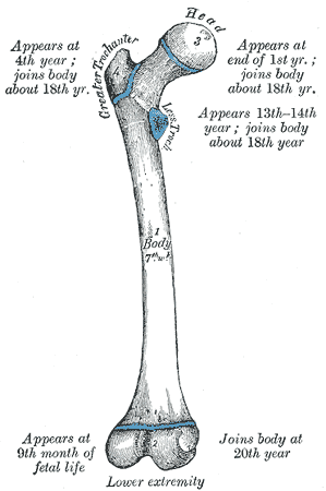
BONE
In the compact bone micrograph at the left, several complete osteons are
visible. In the center of the osteon is the central canal (A) which hold the blood vessels and a nerve. These
canals are surrounded by concentric rings of inorganic matrix, the lamellae (B). Between the lamellae are bone cells, the osteocytes (C) located in lacunae. Nutrients diffuse from cell to
cell through the canaliculi (D).
Location: skeleton
Function: framework, protection are usually considered connective tissue, but
because they differ so substantially from the other tissues in this class, the
phrase "connective tissue proper" is commonly used to exclude those
three. There is also variation in the classification of embryonic connective
tissues; on this page they will be treated as a third and separate category.
Classification
The types of connective tissue proper vary in the type and arrangement of the fibers included and the type of "ground substance" or matrix. The most common cell in these tissues is the fibroblast. (The nuclei stain easily.) The tissues included here are:
AREOLAR CONNECTIVE
|
In the watery matrix (ground substance) observe the nuclei of fibroblasts (A), collagen fibers (B) and elastic fibers (C). Locations: beneath the skin and around blood vessels, muscles and nerves Functions: binds one tissue to another (as skin connects to muscle), protection and nourishment to the organs and structures it binds, and stores "body fluid" |
|
|
|
|
|
|
|
|
|
|
|
|
|
|
|
|
|
|
|
|
|
|
|
|
|
|
|
|
|
|
|
|
|
|
|
|
|
|
|
|
|
|
|
|
|
|
|
|
|
|
|
|
|
|
|
|
|
|
|
|
|
|
|
|
NSE REGULAR CONNECTIVE
The micrograph above is at very low magnification. To the left, at a much higher magnification, the fibroblasts (A) are more clearly observed between the parallel collagenous fibers (B).
Locations:
tendons and ligaments
Functions: strong flexible support
|
This section of aorta shows a tremendous number of elastic fibers (A). The fibroblasts are
not visible. The light pink in this tissue is smooth muscle. |
|||||||||||||||||||||||||||||||||||||||||||||||||||||||||||||||
|
Observe that the reticular fibers (A) form a network or lattice in this spleen tissue. Do not confuse this tissue with the elastic connective tissue seen above which has fibers that are parallel. Locations: spleen, lymph nodes, liver Function: gives support to soft organs
|
ADIPOSE
|
Above observe adipose at a low magnification. The cells appear empty. At the left observe that the nuclus (A) is pushed to the side of the cell giving the cell the appearance of a signet ring. Cells are filled with fat globules (B). Locations/functions: |
|
|
|
|
|
|
|
|
|
|
|
|
|
|
|
|
|
|
|
|
|
|
|
|
|
|
|
|
|
|
|
|
|
|
|
|
|
|
|
|
|
|
|
|
|
|
|
|
|
|
|
|
|
|
|
|
|
|
|
|
|
|
|
|
Connective tissue proper
Areolar (or loose) connective tissue holds organs and epithelia in place, and has a variety of proteinaceous fibres, including collagen and elastin. It is also important in inflammation
· Adipose tissue contains adipocytes, used for cushioning, thermal insulation, lubrication (primarily in the pericardium) and energy storage. [fat]
· Dense connective tissue (or, less commonly, fibrous connective tissue) forms ligaments and tendons. Its densely packed collagen fibres have great tensile strength.
· Reticular connective tissue is a network of reticular fibres (fine collagen, type III) that form a soft skeleton to support the lymphoid organs (lymph nodes, bone marrow, and spleen.)
Specialized connective tissues

· Blood functions in transport. Its extracellular matrix is blood plasma, which transports dissolved nutrients, hormones, and carbon dioxide in the form of bicarbonate. The main cellular component is red blood cells.

· Bone makes up virtually the entire skeleton in adult vertebrates.
· Cartilage makes up virtually the entire skeleton in chondrichthyes. In most other vertebrates, it is found primarily in joints, where it provides cushioning. The extracellular matrix of cartilage is composed primarily of collagen.
http://www.youtube.com/watch?v=WNd6H7l4sOI
|
|
|
|
|
|
|
|
|
|
|
|
|
|
|
|
|
|
|
|
|
|
|
|
|
|
|
|
|
|
|
|
|
|
|
|
|
|
|
|
|
|
|
|
|
|
|
|
|
|
|
|
|
|
|
|
|
|
|
|
|
|
|
|
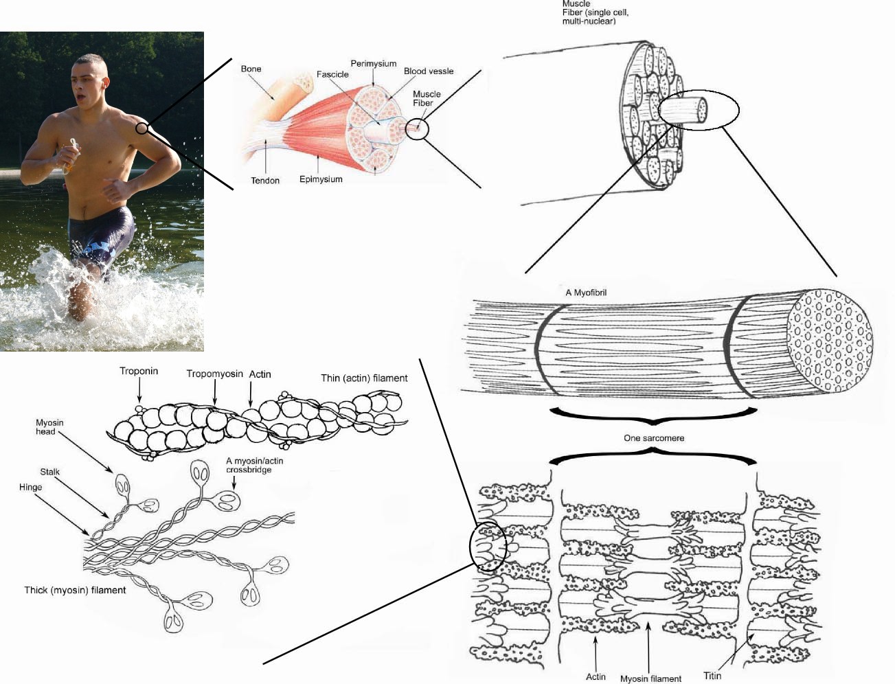
As the name implies, connective tissue serves a "connecting" function. It supports and binds other tissues. Unlike epithelial tissue, connective tissue typically has cells scattered throughout an extracellular matrix.
Epithelial Cell
The cells of connective tissue are embedded in a great amount of extracellular material. This matrix is secreted by the cells. It consists of protein fibers embedded in an amorphous mixture of huge protein-polysaccharide ("proteoglycan") molecules.
Supporting connective tissue
Gives strength, support, and protection to the soft parts of the body.
· Cartilage. Example: the outer ear
· Bone. The matrix of bone contains collagen fibers and mineral deposits. The most abundant mineral is calcium phosphate, although magnesium, carbonate, and fluoride ions are also present.
Binding connective tissue
It binds body parts together.
· Tendons connect muscle to bone. The matrix is principally collagen, and the fibers are all oriented parallel to each other. Tendons are strong but not elastic.
· Ligaments attach one bone to another. They contain both collagen and also the protein elastin. Elastin permits ligaments to be stretched.
|
|
|
|
|
|
|
|
|
|
|
|
|
|
|
|
|
|
|
|
|
|
|
|
|
|
|
|
|
|
|
|
|
|
|
|
|
|
|
|
|
|
|
|
|
|
|
|
|
|
|
|
|
|
|
|
|
|
|
|
|
|
|
|
Fibrous connective tissue
It is distributed throughout the body. It serves as a packing and binding material for most of our organs. Collagen, elastin, and other proteins are found in the matrix.
Fascia is fibrous connective tissue that binds muscle together and binds the skin to the underlying structures. Elastin is a major protein component.
Adipose tissue is fibrous connective tissue in which the cells, called adipocytes, have become almost filled with oil.
Fibrous and binding connective tissue is derived from cells called fibroblasts, which secrete the extracellular matrix.
The extracellular matrix of cartilage and bone is secreted by specialized cells derived from fibroblasts:
· chondroblasts for cartilage;
· osteoblasts for bone.
Composition of the ECM
The ECM of vertebrates is composed of complex mixtures of
· proteins and proteoglycans,
· in the case of bone, mineral deposits.
Proteins
Almost all of the proteins are glycoproteins; that is, have short chains of carbohydrate residues attached to them. (Elastin does not.) A wide variety of collagens. [Link to a page devoted to the collagens.]
· Laminins. Abundant in the basal lamina of epithelia.
· Fibronectin. Binds cells to the ECM.
· Elastins. Provide flexibility to skin, arteries, and lungs. (These are not glycosylated.)
Proteoglycans
Proteoglycans are also glycoproteins but consist of much more carbohydrate than protein; that is, they are huge clusters of carbohydrate chains often attached to a protein backbone.
· The protein backbone of proteoglycans is synthesized, like other secreted proteins, in the endoplasmic reticulum.
· Several sugars are incorporated in proteoglycans. The most abundant one is N-acetylglucosamine (NAG) (the same monomer out of which chitin is made).
· The long chains of sugar residues are attached to serine residues in the protein backbone; that is, they are "O-linked".
· This glycosylation occurs in the Golgi apparatus.
· Sulfate groups are also added to the sugars while in the Golgi apparatus.
· In most cases the completed molecules are then secreted by the cell.
Some examples:
· Chondroitin sulfate
· Heparan sulfate
· Keratan sulfate
· Hyaluronic acid (This one contains literally thousands of NAG residues but does not have a protein component.)
(Their presence in connective tissue like joints accounts for the popularity of N-acetylglucosamine and chondroitin sulfate as dietary supplements for arthritis sufferers.)
Proteoglycans are degraded in lysosomes. A variety of different enzymes are needed. Inherited deficiencies in any one of these produces one of some dozen different types of mucopolysaccharidosis (mucopolysaccharide is the earlier name for proteoglycan).
Syndecan-1
This proteoglycan differs from the others in being retained at the surface of the cell anchored in the plasma membrane as an integral transmembrane protein.
Syndecan-1 binds chemokines (chemotactic cytokines). When epithelia are damaged, these complexes are released and diffuse away forming a chemotactic gradient that attracts neutrophils to the site. Thus syndecan-1 plays a crucial role in inflammation.
Connecting Cells to the ECM
Most normal vertebrate cells cannot survive unless they are anchored to the extracellular matrix. This anchorage dependence is often lost when a cell turns cancerous. (HeLa cells, for example, are among the few types of vertebrate cell that can be grown in liquid culture.)
Cells attach to the ECM by means of transmembrane glycoproteins called integrins.
· The extracellular portion of integrins binds to various types of ECM proteins:
· collagens
· laminins
· fibronectin
· The intracellular portion binds to the actin filaments of the cytoskeleton.
|
|
|
|
|
|
|
|
|
|
|
|
|
|
|
|
|
|
|
|
|
|
|
|
|
|
|
|
|
|
|
|
|
|
|
|
|
|
|
|
|
|
|
|
|
|
|
|
|
|
|
|
|
|
|
|
|
|
|
|
|
|
|
|
Cancer Metastasis
Cancers begin as a primary tumor. At some point, however, cells break away from the primary tumor and - traveling in blood and lymph - establish metastases in other locations of the body. Metastasis is what usually kills the patient.
In order to enter (and exit) the blood or lymph, cancer cells must pass through a basement membrane. They are able to do so by secreting proteinases (including serine proteases) that digest a path for them.
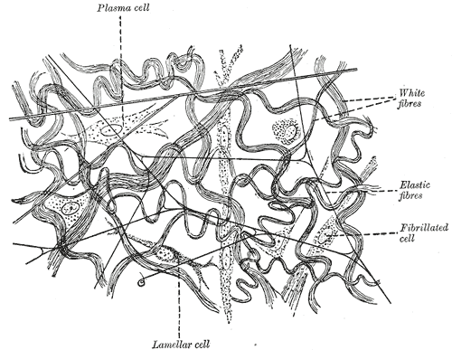
Loose Connective Tissue
In vertebrates, the most common type of
connective tissue is loose connective tissue. It holds organs in place and attaches
epithelial tissue to other underlying tissues.
Loose connective tissue is named based on the "weave" and type of its
constituent fibers. There are three main types:
Collagenous fibers
are made of collagen and consist of bundles of fibrils that are coils of collagen molecules.
Elastic Fibers
Elastic fibers are made of elastin and are "stretchable."
Reticular Fibers
Reticular fibers join connective tissues to other tissues.
|
|
|
|
|
|
|
|
|
|
|
|
|
|
|
|
|
|
|
|
|
|
|
|
|
|
|
|
|
|
|
|
|
|
|
|
|
|
|
|
|
|
|
|
|
|
|
|
|
|
|
|
|
|
|
|
|
|
|
|
|
|
|
|
Fibrous Connective Tissue
Another type of connective tissue is fibrous connective tissue which is found in tendons and ligaments. Fibrous connective tissue is composed of large amounts of closely packed collagenous fibers.
Specialized Connective Tissues
Adipose tissue is a form of loose connective tissue that stores fat.
Cartilage is a form of fibrous connective tissue that is composed
of closely packed collagenous fibers in a rubbery gelatinous substance called
chondrin. The skeletons of sharks and human embryos are composed of cartilage.
Cartilage also provides flexible support for certain structures in adult humans
including the nose, trachea and ears.
Bone is a type of mineralized connective tissue that
contains collagen and calcium phosphate, a mineral crystal. Calcium phosphate
gives bone its firmness.
Interestingly enough, blood is considered to be a type of connective tissue. Even
though it has a different function in comparison to other connective tissues it
does have an extracellular matrix. The matrix is the plasma and erythrocytes, leukocytes and platelets are suspended in the plasma.
|
|
|
|
Human Blood Cells |
|
Embryonic connective tissues
· Mesenchymal connective tissue
·


Fiber types
Fiber types as follows:
·

·
Disorders of connective tissue
Various connective tissue conditions have been identified; these can be both inherited and environmental.
· Marfan syndrome - a genetic disease causing abnormal fibrillin.
|
|
|
|
|
|
|
|
|
|
|
|
|
|
|
|
|
|
|
|
|
|
|
|
|
|
|
|
|
|
|
|
|
|
|
|
|
|
|
|
|
|
|
|
|
|
|
|
|
|
|
|
|
|
|
|
|
|
|
|
|
|
|
|

· Scurvy - caused by a dietary deficiency in vitamin C, leading to abnormal collagen.
· Ehlers-Danlos syndrome - deficient type III collagen- a genetic disease causing progressive deterioration of collagens, with different EDS types affecting different sites in the body, such as joints, heart valves, organ walls, arterial walls, etc.
·

A representation of a condensed eukaryotic chromosome, as seen during cell division.
· Loeys-Dietz syndrome - a genetic disease related to Marfan syndrome, with an emphasis on vascular deterioration.
· Systemic lupus erythematosus - a chronic, multisystem, inflammatory disorder of probable autoimmune etiology, occurring predominantly in young women.
· Osteogenesis imperfecta (brittle bone disease) - caused by insufficient production of good quality collagen to produce healthy, strong bones.
· Fibrodysplasia ossificans progressiva - disease of the connective tissue, caused by a defective gene which turns connective tissue into bone.
· Spontaneous pneumothorax - collapsed lung, believed to be related to subtle abnormalities in connective tissue.
Pneumothorax

· Sarcoma - a neoplastic process originating within connective tissue.
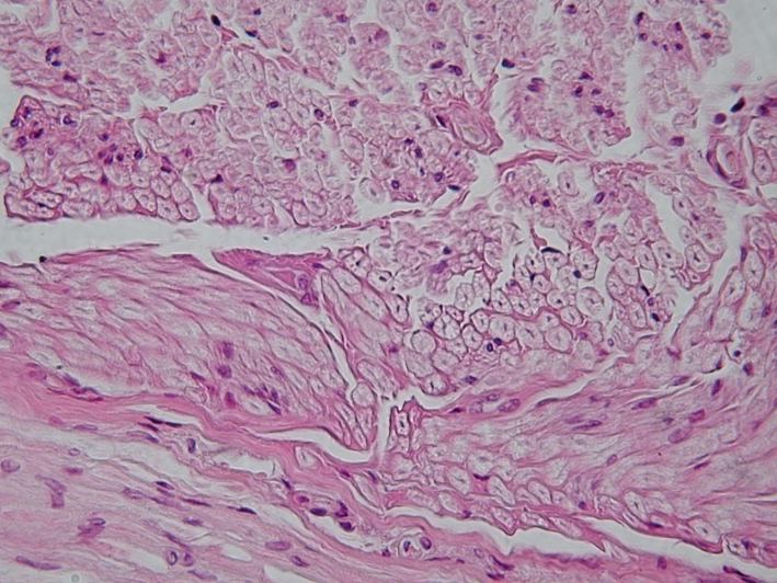
Staining of connective tissue
For microscopic viewing, the majority of the connective tissue staining techniques color tissue fibers in contrasting shades. Collagen may be differentially stained by any of the following techniques:
· Van Gieson's stain
· Masson's Trichrome stain
·

Microscopy of keratin filaments inside cells.
· Mallory's Aniline Blue stain
· Azocarmine stain
· Krajian's Aniline Blue stain
|
|
|
|
|
|
|
|
|
|
|
|
|
|
|
|
|
|
|
|
|
|
|
|
|
|
|
|
|
|
|
|
|
|
|
|
|
|
|
|
|
|
|
|
|
|
|
|
|
|
|
|
|
|
|
|
|
|
|
|
|
|
|
|
|
|
|
|
|
|
|
|
|
|
|
|
|
|
|
|
|
|
|
|
|
|
|
|
|
|
|
|
|
|
|
|
|
|
|
|
|
|
|
|
|
|
|
|
|
|
|
|
|
|
|
|
|
|
|
|
|
|
|
|
|
|
|
|
|
|
|
|
|
|
|
|
|
|
|
|
|
|
|
|
|
|
|
|
|
|
|
|
|
|
|
|
|
|
|
|
|
|
|
|
|
|
|
|
|
|
|
|
|
|
|
|
|
|
|
|
|
|
|
|
|
|
|
|
|
|
|
|
|
|
|
|
|
|
|
|
|
|
|
|
|
|
|
|
|
|
|
|
|
|
|
|
|
|
|
|
|
|
|
|
|
|
|
|
|
|
|
|
|
|
|
|
|
|
|
|
|
|
|
|
|
|
|
|
|
|
|
|
|
|
|
|
|
|
|
|
|
|
|
|
|
|
|
|
|
|
|
|
|
|
|
|
|
|
|
|
|
|
|
|
|
|
|
|
|
|
|
|
|
|
|
|
|
|
|
|
|
|
|
|
|
|
|
|
|
|
|
|
|
|
|
|
|
|
|
|
|
|
|
|
|
|
|
|
|
|
|
|
|
|
|
|
|
|
|
|
|
|
|
|
|
|
|
|
|
|
|
|
|
|
|
|
|
|
|
|
|
|
|
|
|
|
|
|
|
|
|
|
|
|
|
|
|
|
|
|
|
|
|
|
|
|
|
|
|
|
|
|
|
|
|
|
|
|
|
|
|
|
|
|
|
|
|
|
|
|
|
|
|
|
|
|
|
|
|
|
|
|
|
|
|
|
|
|
|
|
|
|
|
|
|
|
|
|
|
|
|
|
|
|
|
|
|
|