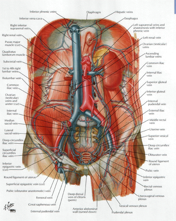
1. Inferior vena cava system. System of portal vein. Foetal circulation. Anastomoses between the portal and
general systemic circulation
2. Autonomic
nervous system.Central and peripheral parts of sympathetic and parasympathetic autonomic
nervous system
3. Preparation
of vessels and nerves of cavities
Lesson # 28
Theme
1. System of inferior vena cava. System of portal vein. Foetal
circulation. Anastomoses between the portal and general systemic
circulation
Venae cavae are the two largest veins in the body. These blood vessels carry de-oxygenated
blood from various regions of the body to the right atrium of the heart. As the de-oxygenated
blood is returned to the heart and continues to flow through the cardiac cycle,
it is transported to the lungs where it becomes oxygenated. The blood then
travels back to the heart and is pumped out to the rest of the body via the aorta. Oxygen depleted blood
is returned to the heart again via the venae cavae.
The superior vena cava is located in the upper chest region and is
formed by the joining of the brachiocephalic veins.
It is bordered by heart structures such as the aorta and pulmonary artery.
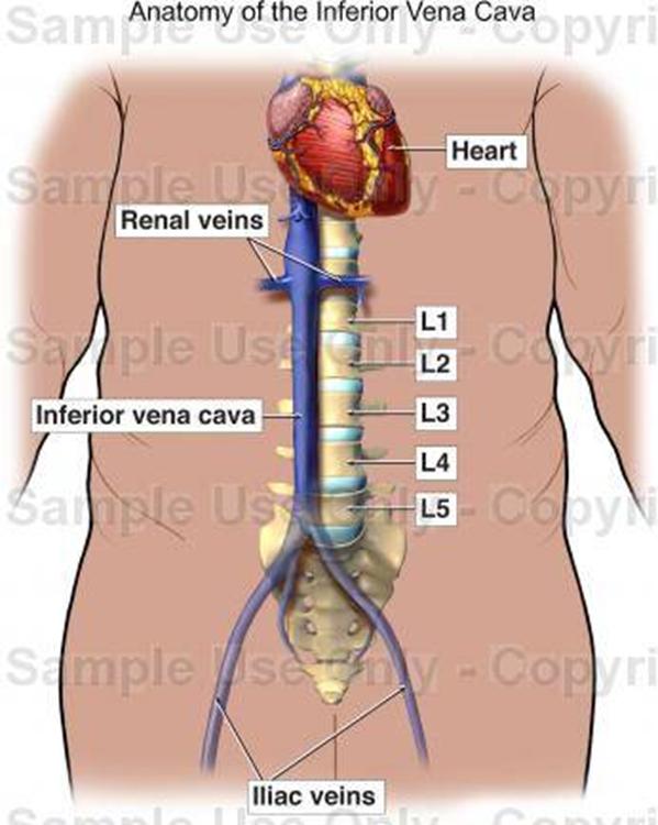
The inferior vena cava is formed by the joining of the common iliac veins
which meet a little below the small of the back. The inferior vena cava travels
along the spine and transports blood from the lower extremities of the body to the
posterior region of the right atrium.
Function of the Venae Cavae
·
Superior Vena Cava: Brings de-oxygenated blood from
the head, neck, arm and chest regions of the body to the right atrium.
·
Inferior Vena Cava: Brings de-oxygenated blood from
the lower body regions (legs, back, abdomen and pelvis) to the right atrium.
The inferior vena cava
(or IVC), also known as the posterior vena cava, is the large vein that carries
de-oxygenated blood from the lower half of
the body into the right atrium of the heart.
It is posterior to the abdominal cavity and runs alongside of the vertebral column
on its right side (i.e. it is a retroperitoneal
structure). It enters the right atrium at the lower right, back side of the heart.
The IVC is formed by the joining of the left and right common iliac veins
and brings blood into the right atrium of the heart. It also anastomoses with the azygos vein system (which
runs on the rght side of the vertebral column) and
venous plexuses next to the spinal cord.

Because the IC is not centrally located, there are
some asymmetries in drainage patterns. The gonadal veins
and suprarenal veins
drain into the IVC on the right side, but into the renal vein
on the left side, which in turn drains into the IVC. By contrast, all the lumbar veins
and hepatic veins
usually drain directly into the IVC.
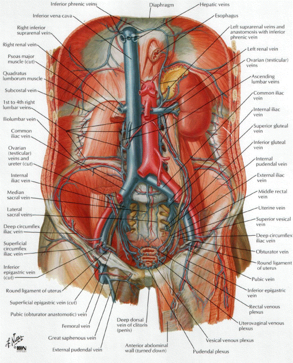
The tributaries of Inferior vena cava can be remembered using the mnemonic, "I Like To Rise So High", for Illiac
vein (common), Lumbar vein, Testicular vein, Renal vein, Suprarenal vein and
Hepatic vein.[2]
Note that the vein that carries de-oxygenated blood from the upper half
of the body is the superior vena cava

Inferior vena cava starts on level IV-V lumbar
vertebrae by the confluence of left common iliac vein and right common iliac
vein, to the right and beneath from bifurcation of aorta. It passes through
special foramen in centrum tendineum of diaphragm
into mediastinum and empties into right atrium.
There are parietal and visceral influxes of inferior vena cava.

The venæ cavæ and azygos veins, with their tributaries.
Parietal tributaries of inferior
vena cava:
·
lumbar veins are
3-4 pairs, which collect blood from areas according with ramification of lumbar
arteries, they anastomose by right and left ascending lumbar veins;
inferior phrenic veins collect blood from areas according with ramification same name
arteries.


·
Follow veins are the visceral tributaries of inferior vena
cava:
·
in
male - right testicular vein starts from posterior testicle margin.
Testicular vein forms pampiniform plexus
which enters to composition of spermatic cord. Left testicular vein
(also left ovaricа vein in famile)
empties by right angle into left renal vein;
·
in famile -
right ovaric
·
vein begins from
ovary hilus;
·
renal veins,
pair, pass from kidney hilus and, anastomosing with
lumbar veins, emptiy into inferior vena cava between
lumbar vertebrae first and second;
·
right
suprarenal vein, exits
from hilus of adrenal gland. Left
suprarenal vein falls into left renal vein;
·
hepatic
veins (3-4) veins fall into inferior vena cava in area of
same name sulcus in liver.
The inferior vena cava (v. cava inferior),
returns to the heart the blood from the parts below the diaphragm. It is formed
by the junction of the two common iliac veins, on the right side of the fifth
lumbar vertebra. It ascends along the front of the vertebral column, on the
right side of the aorta, and, having reached the liver, is continued in a
groove on its posterior surface. It then perforates the diaphragm between the
median and right portions of its central tendon; it subsequently inclines forward
and medialward for about
Relations.—The abdominal
portion of the inferior vena cava is in relation in front, from
below upward, with the right common iliac artery, the mesentery, the right
internal testicular artery, the inferior part of the duodenum, the pancreas,
the common bile duct, the portal vein, and the posterior surface of the liver;
the last partly overlaps and occasionally completely surrounds it; behind,
with the vertebral column, the right Psoas major, the right crus of the
diaphragm, the right inferior phrenic, suprarenal, renal and lumbar arteries,
right sympathetic trunk and right celiac ganglion, and the medial part of the
right suprarenal gland; on the right side, with the right kidney and
ureter; on the left side, with the aorta, right crus of the diaphragm,
and the caudate lobe of the liver.
The thoracic portion is only about
Peculiarities.—In Position.—This
vessel is sometimes placed on the left side of the aorta, as high as the left
renal vein, and, after receiving this vein, crosses over to its usual position
on the right side; or it may be placed altogether on the left side of the
aorta, and in such a case the abdominal and thoracic viscera, together with the
great vessels, are all transposed.
Point of Termination.—Occasionally the inferior vena
cava joins the azygos vein, which is then of large
size. In such cases, the superior vena cava receives the whole of the blood
from the body before transmitting it to the right atrium, except the blood from
the hepatic veins, which passes directly into the right atrium.
Tributaries.—The
inferior vena cava receives the following veins:
Lumbar.
Renal.
Inferior Phrenic.
Right Spermatic or Ovarian.
Suprarenal.
Hepatic.
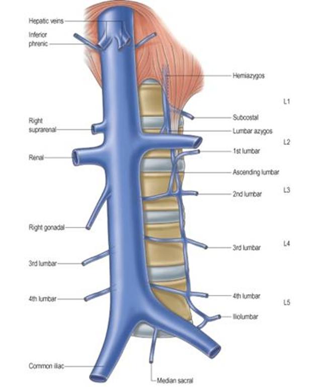
The Lumbar Veins (vv. lumbales)
four in number on each side, collect the blood by dorsal tributaries
from the muscles and integument of the loins, and by abdominal tributaries from
the walls of the abdomen, where they communicate with the epigastric
veins. At the vertebral column, they receive veins from the vertebral plexuses,
and then pass forward, around the sides of the bodies of the vertebræ, beneath the Psoas major, and end in the
back part of the inferior cava. The left lumbar veins are longer than the
right, and pass behind the aorta. The lumbar veins are connected together by a
longitudinal vein which passes in front of the transverse processes of the
lumbar vertebræ, and is called the ascending
lumbar; it forms the most frequent origin of the corresponding azygos or hemiazygos vein, and
serves to connect the common iliac, iliolumbar, and azygos or hemiazygos veins of its
own side of the body.


The Testicular veins (vv. spermaticæ)
emerge from the back of the testis, and receive tributaries from the
epididymis; they unite and form a convoluted plexus, called the pampiniform plexus, which constitutes the
greater mass of the spermatic cord; the vessels composing this plexus are very
numerous, and ascend along the cord, in front of the ductus
deferens. Below the subcutaneous inguinal ring they unite to form three or four
veins, which pass along the inguinal canal, and, entering the abdomen through
the abdominal inguinal ring, coalesce to form two veins, which ascend on the
Psoas major, behind the peritoneum, lying one on either side of the internal
testicular artery. These unite to form a single vein, which opens on the right
side into the inferior vena cava, at an acute angle; on the left side into the
left renal vein, at a right angle. The testicular veins are provided with
valves. 107 The left testicular vein passes behind the iliac colon, and is thus
exposed to pressure from the contents of that part of the bowel.
The Ovarian Veins (vv. ovaricæ)
correspond with the spermatic in the male; they form a plexus in the broad
ligament near the ovary and uterine tube, and communicate with the uterine
plexus. They end in the same way as the testicular veins in the male. Valves
are occasionally found in these veins. Like the uterine veins,
they
become much enlarged during pregnancy.

Testicular
veins.
The Renal Veins (vv. renales)
are of large size, and placed in front of the renal arteries. The left is
longer than the right, and passes in front of the aorta, just below the origin
of the superior mesenteric artery. It receives the left testicular and left
inferior phrenic veins, and, generally, the left suprarenal vein. It opens into
the inferior vena cava at a slightly higher level than the right.
The Suprarenal Veins (vv. suprarenales)
are two in number: the right ends in the inferior vena cava; the left, in the
left renal or left inferior phrenic vein.
The Inferior Phrenic Veins (vv. phrenicæ inferiores)
follow the course of the inferior phrenic arteris;
the right ends in the inferior vena cava; the left is often represented by two
branches, one of which ends in the left renal or suprarenal vein, while the
other passes in front of the esophageal hiatus in the
diaphragm and opens into the inferior vena cava.
The Hepatic Veins (vv. hepaticæ)
commence in the substance of the liver, in the terminations of the portal vein
and hepatic artery, and are arranged in two groups, upper and lower. The upper
group usually consists of three large veins, which converge toward the
posterior surface of the liver, and open into the inferior vena cava, while
that vessel is situated in the groove on the back part of the liver. The veins
of the lower group vary in number, and are of small size; they come from
the right and caudate lobes. The hepatic veins run singly, and are in direct
contact with the hepatic tissue. They are
destitute of valves.

The
portal vein and its tributaries.
Vena
portae hepatis
is situated in thickness of hepatoduodenal
ligament between ductus choledochus
and proper hepatic artery (formula of their position DVA – from right to left).
It originates behind head of pancreas by the confluence of superior,
inferior mesenteric veinc and splenic
vein. It collects venous blood from odd organs of abdominal cavity, except
liver. Vena portae receives cystic vein,
right and left gastric veins and prepyloric
vein closely to liver hilus. Paraumbilical
veins fall into portal vein in liver hilus.
There
are cava-caval and porto-caval anastomoses between systems of portal vein,
superior and inferior vanea cavae
(see table on the next page).

The portal system includes all the veins which drain the blood
from the abdominal part of the digestive tube (with the exception of the lower
part of the rectum) and from the spleen, pancreas, and gall-bladder. From these
viscera the blood is conveyed to the liver by the portal vein. In the
liver this vein ramifies like an artery and ends in capillary-like vessels termed
sinusoids, from which the blood is conveyed to the inferior vena cava by
the hepatic veins. From this it will be seen that the blood of the portal
system passes through two sets of minute vessels, viz., (a) the
capillaries of the digestive tube, spleen, pancreas, and gall-bladder; and (b)
the sinusoids of the liver. In the adult the portal vein and its tributaries
are destitute of valves; in the fetus and for a short
time after birth valves can be demonstrated in the tributaries of the portal
vein; as a rule they soon atrophy and disappear, but in some subjects they
persist in a degenerate form.

The portal vein (vena portæ)
is about
venosus).

Tributaries.—The
tributaries of the portal vein are:
Lienal.
Pyloric.
Cystic.
Coronary.
Parumbilical.
The Lienal Vein (v.
lienalis; splenic vein) commences by five
or six large branches which return the blood from the spleen. These unite to
form a single vessel, which passes from left to right, grooving the upper and
back part of the pancreas, below the lineal artery, and ends behind the neck of
the pancreas by uniting at a right angle with the superior mesenteric to form
the portal vein. The lienal vein is of large size,
but is not tortuous like the artery.

Tributaries.—The
lineal vein receives the short gastric veins, the left gastroepiploic
vein, the pancreatic veins, and the inferior mesenteric veins.
The short gastric veins (vv. gastricæ
breves), four or five in number, drain the fundus
and left part of the greater curvature of the stomach, and pass between the two
layers of the gastrolienal ligament to end in the lienal vein or in one of its large tributaries.
The left gastroepiploic vein
(v. gastroepiploica sinistra)
receives branches from the antero-superior and postero-inferior surfaces of the stomach and from the
greater omentum; it runs from right to left along the
greater curvature of the stomach and ends in the commencement of the lienal vein.

The pancreatic veins (vv. pancreaticæ)
consist of several small vessels which drain the body and tail of the pancreas,
and open into the trunk of the lienal vein.
The inferior mesenteric vein (v.
mesenterica inferior) returns blood from the
rectum and the sigmoid, and descending parts of the colon. It begins in
the rectum as the superior hemorrhoidal vein,
which has its origin in the hemorrhoidal plexus, and
through this plexus communicates with the middle and inferior hemorrhoidal veins. The superior hemorrhoidal
vein leaves the lesser pelvis and crosses the left common iliac vessels with
the superior hemorrhoidal artery, and is continued
upward as the inferior mesenteric vein. This vein lies to the left of its
artery, and ascends behind the peritoneum and in front of the left Psoas major;
it then passes behind the body of the pancreas and opens into the lienal vein; sometimes it ends in the angle of union of the
lienal and superior mesenteric veins.

Tributaries.—The
inferior mesenteric vein receives the sigmoid veins from the sigmoid
colon and iliac colon, and the left colic vein from the descending colon
and left colic flexure.
The Superior Mesenteric Vein (v. mesenterica superior) returns the blood from the small
intestine, from the cecum, and from the ascending and transverse portions of the
colon. It begins in the right iliac fossa by the union of the veins which drain
the terminal part of the ileum, the cecum, and vermiform process, and ascends
between the two layers of the mesentery on the right side of the superior
mesenteric artery. In its upward course it passes in front of the right ureter,
the inferior vena cava, the inferior part of the duodenum, and the lower
portion of the head of the pancreas. Behind the neck of the pancreas it unites
with the lienal vein to form the portal vein.

Tributaries.—Besides
the tributaries which correspond with the branches of the superior mesenteric
artery, viz., the intestinal, ileocolic, right
colic, and middle colic veins, the superior mesenteric vein is
joined by the right gastroepiploic and pancreaticoduodenal veins.
The right gastroepiploic vein
(v. gastroepiploica dextra)
receives branches from the greater omentum and from
the lower parts of the antero-superior and posteroinferior surfaces of the stomach; it runs from left
to right along the greater curvature of the stomach between the two layers of
the greater omentum.
The pancreaticoduodenal
veins (vv. pancreaticoduodenales)
accompany their corresponding arteries; the lower of the two frequently joins
the right gastroepiploic vein.
The Coronary Vein (v. coronaria
ventriculi; gastric vein) derives tributaries
from both surfaces of the stomach; it runs from right to left along the lesser
curvature of the stomach, between the two layers of the lesser omentum, to the esophageal
opening of the stomach, where it receives some esophageal
veins. It then turns backward and passes from left to right behind the omental bursa and ends in the portal vein.
The Pyloric Vein is of small size, and runs from left
to right along the pyloric portion of the lesser curvature of the stomach,
between the two layers of the lesser omentum, to end
in the portal vein.
The Cystic Vein (v. cystica)
drains the blood from the gall-bladder, and, accompanying the cystic duct,
usually ends in the right branch of the portal vein.
Parumbilical Veins (vv. parumbilicales).—In the course of the ligamentum teres of the liver and of the middle umbilical ligament,
small veins (parumbilical) are found which establish
an anastomosis between the veins of the anterior abdominal wall and the portal,
hypogastric, and iliac veins. The best marked of
these small veins is one which commences at the umbilicus and runs backward and
upward in, or on the surface of, the ligamentum teres between the layers of the falciform
ligament to end in the left portal vein.
Collateral venous circulation to relieve portal obstruction
in the liver may be effected by communications between (a) the gastric
veins and the esophageal veins which often project as
a varicose bunch into the stomach, emptying themselves into the hemiazygos vein; (b) the veins of the colon and
duodenum and the left renal vein; (c) the accessory portal system of Sappey, branches of which pass in the round and falciform ligaments (particularly the latter) to unite with
the epigastric and internal mammary veins, and
through the diaphragmatic veins with the azygos; a
single large vein, shown to be a parumbilical vein,
may pass from the hilus of the liver by the round
ligament to the umbilicus, producing there a bunch of prominent varicose veins
known as the caput medusæ; (d)
the veins of Retzius, which connect the intestinal
veins with the inferior vena cava and its retroperitoneal branches; (e)
the inferior mesenteric veins, and the hemorrhoidal
veins that open into the hypogastrics; (f)
very rarely the ductus venosus
remains patent, affording a direct connection between the portal vein and the
inferior vena cava.
Anastomoses between the superior and inferior venae
cavae systems
|
System |
Veins |
Veins |
Position of
anastomose |
|
V. cava
superior And v. cava
inferior |
v. epigastrica superior (tributary of the internal thoracic
vein) and v. Thoracoepigastrica (tributary of the subclavian vein) |
v. epigastrica inferior (tributary of the external iliac
vein) and v. Epigastrica superficialis
(tributary of the femoral vein) |
In anterior
abdominal wall round the navel |
|
V. cava
superior And v. Cava
inferior |
vv. azygos and hemiazygos |
vv. lumbales |
On
posterior abdominal wall |
|
V. cava
superior And v. Cava
inferior |
Rr.
spinales (tributary of the vv. Intercostales
posteriores) |
Rr.
spinales (tributary of the vv. Lumbales)
|
Form
internal and external vertebral plexus |
Anastomoses between the superior and inferior
venae cavae
and
portal vein systems
|
V. cava
superior and v. Portae |
v. epigastrica superior (tributary of the internal thoracic
vein) |
Vv. paraumbilicales |
In anterior
abdominal wall round the navel |
|
V. cava superior
and v. Portae |
Vv. esophageales (tributary of the azygos
vein) |
v. gastrica sinistra |
Near
gastric cardia |
|
V. cava
inferior and v. Portae |
v. epigastrica inferior (tributary of the external iliac
vein) |
Vv. paraumbilicales |
In anterior
abdominal wall |
|
V. cava
inferior and v. Portae |
V. rectalis media (tributary of the internal iliac vein) |
V. rectalis superior (tributary of the inferior mesenteric
vein) |
Plexus venosus rectalis |
|
V. cava
inferior and v. Portae |
Vv. lumbales |
Vv. mesenterica superior and inferior |
In
thickness of ascending and descending colon |
Circulatory system of the foetus
has a row of peculiarities that differ from adult one:
-
arterial blood reaches the foetus
through umbilical vein from placenta;
-
exclusive of umbilical vein, a blood
in vessels is mixed;
-
venous (Аranti)
duct functions between umbilical and inferior vena cava by
veins;
-
blood from inferior vena cava gets
from right atrium through the ovale foramen
into left atrium;
-
pulmonary circulation does not function;
-
arterial (Botalova)
duct functions between aortic arch and pulmonary trunk,
through the which blood from pulmonary blood circle passes in systemic
circulation;
-
more
oxygenated blood supplies head, neck, upper limbs and superior part of torso.
Inferior part of trunk and lower limbs supplied by mixed blood, which is
insufficiently saturated by oxygen, that's why these body portions of foetus
fall behind in development in compare of head and upper part of torso.
After
birth breath starts and pulmonary circulation begins to function. Umbilical
vessels overgrow in 6-7 days, Botali duct - in 9-10 days
and oval foramen in interatrial wall – in 30 days
after birth

Peculiarities
in the Vascular System in the Fetus The chief peculiarities of the fetal
heart are the direct communication between the atria through the foramen ovale, and the large size of the valve of the inferior vena
cava. Among other peculiarities the following may be noted. (1) In early fetal life the heart lies immediately below the mandibular
arch and is relatively large in size. As development proceeds it is gradually
drawn within the thorax, but at first it lies in the middle line; toward the
end of pregnancy it gradually becomes oblique in direction. (2) For a time the
atrial portion exceeds the ventricular in size, and
the walls of the ventricles are of equal thickness: toward the end of fetal life the ventricular portion becomes the larger and
the wall of the left ventricle exceeds that of the right in thickness. (3) Its
size is large as compared with that of the rest of the body, the proportion at
the second month being 1 to 50, and at birth, 1 to 120, while in the adult the
average is about 1 to 160.
The foramen ovale, situated
at the lower part of the atrial septum, forms a free communication between the
atria until the end of fetal life. A septum (septum
secundum) grows down from the upper wall of the
atrium to the right of the primary septum in which the foramen ovale is situated; shortly after birth it fuses with the
primary septum and the foramen ovale is obliterated.
The valve of the inferior vena cava serves to direct
the blood from that vessel through the foramen ovale
into the left atrium.
The peculiarities in the arterial system of the fetus are the communication between the pulmonary artery
and the aorta by means of the ductus arteriosus, and the continuation of the hypogastric
arteries as the umbilical arteries to the placenta.
The ductus arteriosus is a short tube, about
The hypogastric arteries
run along the sides of the bladder and thence upward on the back of the
anterior abdominal wall to the umbilicus; here they pass out of the abdomen and
are continued as the umbilical arteries in the umbilical cord to the
placenta. They convey the fetal blood to the
placenta.
The peculiarities in the venous system of the fetus are the communications established between the
placenta and the liver and portal vein, through the umbilical vein; and between
the umbilical vein and the inferior vena cava through the ductus
venosus.
Fetal Circulation
The abdominal
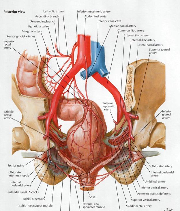
portion of the sympathetic trunk Celiacplexus continues
around abdominal aorta, formingabdominal
aortic plexus. It innervates kidney, suprarenal glands,ureters and
testicles (ovaries).
Theceliac Plexus (Plexus Cœliacus; Solar
Plexus) (
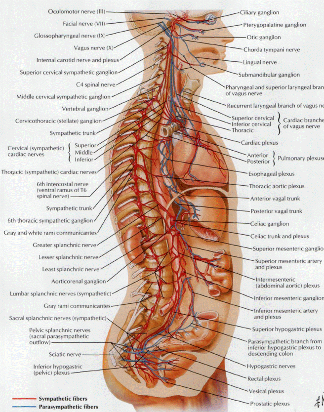

Theceliac
ganglia with the sympathetic plexuses of the abdominal viscera radiatingfrom
the ganglia.


Lesser
curvature of stomach
a. hepatica propria (rr. Dexter et sinister)
The
celiac artery and its branches; the stomach has been raised and the peritoneum
removed.


The
superior mesenteric artery and its branches.
intestini tenuis) arise from the convex side of the superior
mesenteric artery. They are usually from twelve to fifteen in number, and are
distributed to the jejunum and ileum. They run nearly parallel with one another
between the layers of the mesentery, each vessel dividing into two branches,
which unite with adjacent branches, forming a series of arches, the convexities
of which are directed toward the intestine. The Ileocolic Artery (a. ileocolica) is the lowest branch arising
from the concavity of the superior mesenteric artery. It passes downward and to
the right behind the peritoneum toward the right iliac fossa, where it divides
into a superior and an inferior branch; the inferior anastomoses with the end
of the superior mesenteric artery, the superior with the right colic artery.