1. Common and external carotid arteries.
2. ANTERIOR, MEDIAL
AND POSTERIOR branches OF External carotid artery
3. Terminal branches of external carotid artery.
Lesson # 25
Brachiocephalic
trunk begins from aortic arch on level of right II costal
cartilage. It passes upward and to the right of and on level of right sternо-clavicular joint divides into two terminal branches
- right common carotid and right subclavianу
arteries.
Brachiocephalic trunk is the largest branch of the arch of the aorta, and is from 4 to 5 cm. in length. It arises,
on a level with the upper border of the second right costal cartilage, from the
commencement of the arch of the aorta, on a plane anterior to the origin of the
left carotid; it ascends obliquely upward, backward, and to the right to the
level of the upper border of the right sternoclavicular
articulation, where it divides into the right common carotid and right subclavian arteries.

Relations.—Anteriorly, it
is separated from the manubrium sterni by the Sternohyoideus and
Sternothyreoideus, the remains of the thymus, the left innominate and right
inferior thyroid veins which cross its root, and sometimes the superior cardiac
branches of the right vagus. Posterior to it is the trachea, which it
crosses obliquely. On the right side are the right innominate vein, the
superior vena cava, the right phrenic nerve, and the pleura; and on the left
side, the remains of the thymus, the origin of the left common carotid
artery, the inferior thyroid veins, and the trachea.

The arch of the aorta, and its branches.

Branches.—The brachiocephalic trunk usually
gives off no branches; but occasionally a small branch, the thyreoidea ima,
arises from it. Sometimes it gives off a thymic or bronchial branch.
The thyreoidea ima (a. thyreoidea ima) ascends
in front of the trachea to the lower part of the thyroid gland, which it
supplies. It varies greatly in size, and appears to compensate for deficiency
or absence of one of the other thyroid vessels. It occasionally arises from the
aorta, the right common carotid, the subclavian or the internal mammary.
Point of Division.—The brachiocephalic
trunk sometimes divides above the level of the sternoclavicular joint, less
frequently below it.
Position.—When the aortic arch is on the right side, the
innominate is directed to the left side of the neck.

Superficial
dissection of the right side of the neck, showing the carotid and subclavian
arteries.

Collateral Circulation.—Allan Burns demonstrated, on the
dead subject, the possibility of the establishment of the collateral
circulation after ligature of the brachiocephalic trunk, by tying and dividing
that artery. He then found that “Even coarse injection, impelled into the
aorta, passed freely by the anastomosing branches into the arteries of the
right arm, filling them and all the vessels of the head completely.” The branches by which this circulation would be carried on
are very numerous; thus, all the communications across the middle line between
the branches of the carotid arteries of opposite sides would be available for
the supply of blood to the right side of the head and neck; while the
anastomosis between the costocervical of the subclavian and the first aortic
intercostal (see infra on the collateral circulation after obliteration
of the thoracic aorta) would bring the blood, by a free and direct course, into
the right subclavian. The numerous connections, also, between the intercostal
arteries and the branches of the axillary and internal mammary arteries would,
doubtless, assist in the supply of blood to the right arm, while the inferior
epigastric from the external iliac would, by means of its anastomosis with the
internal mammary, compensate for any deficiency in the vascularity of the wall
of the chest.
Common
carotid artery passes behind sternocleidomastoid
muscle upward on front of transverse processes of cervical vertebrae and does
not give off any branches. On the level of upper edge of thyroid cartilage common
carotid artery divides into external carotid artery and internal carotid
artery. This place called bifurcation of carotid artery. There are carotid
sinus and carotid glomus here.
The Arteries of the Head and Neck.
The principal arteries of supply to the head and neck are the two common
carotids; they ascend in the neck and each divides into two branches, viz.,
(1) the external carotid, supplying the exterior of the head, the face,
and the greater part of the neck; (2) the internal carotid, supplying to
a great extent the parts within the cranial and orbital cavities.
The Common Carotid Artery (A. Carotis Communis)—The common carotid arteries differ in length and in their mode
of origin. The right begins at the bifurcation of the brachiocephalic
trunk behind the sternoclavicular joint and is confined to the neck. The left
springs from the highest part of the arch of the aorta to the left of, and on a
plane posterior to the brachiocephalic trunk, and therefore consists of a
thoracic and a cervical portion.
The thoracic portion of the left common carotid artery
ascends from the arch of the aorta through the superior mediastinum to the
level of the left sternoclavicular joint, where it is continuous with the
cervical portion.
1. Relations.—In front, it is
separated from the manubrium sterni by the Sternohyoideus and
Sternothyreoideus, the anterior portions of the left pleura and lung, the left
innominate vein, and the remains of the thymus; behind, it lies on the
trachea, esophagus, left recurrent nerve, and thoracic duct. To its right
side below is the brachiocephalic trunk, and above, the trachea, the
inferior thyroid veins, and the remains of the thymus; to its left side
are the left vagus and phrenic nerves, left pleura, and lung. The left
subclavian artery is posterior and slightly lateral to it.
The cervical portions of the common carotids resemble
each other so closely that one description will apply to both. Each vessel
passes obliquely upward, from behind the sternoclavicular articulation, to the
level of the upper border of the thyroid cartilage, where it divides into the
external and internal carotid arteries.

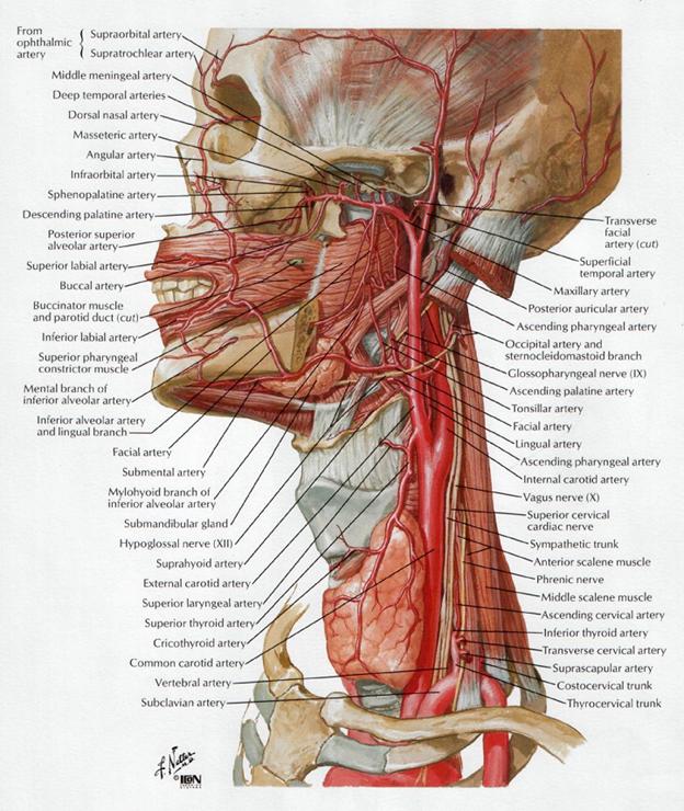
The arteries of the face and scalp.
At the lower part of the neck the two common carotid
arteries are separated from each other by a very narrow interval which contains
the trachea; but at the upper part, the thyroid gland, the larynx and pharynx
project forward between the two vessels. The common carotid artery is contained
in a sheath, which is derived from the deep cervical fascia and encloses also
the internal jugular vein and vagus nerve, the vein lying lateral to the
artery, and the nerve between the artery and vein, on a plane posterior to
both. On opening the sheath, each of these three structures is seen to have a
separate fibrous investment.

Relations.—At the lower part of
the neck the common carotid artery is very deeply seated, being covered by
the integument, superficial fascia, Platysma, and deep cervical fascia, the
Sternocleidomastoideus, Sternohyoideus, Sternothyreoideus, and Omohyoideus; in
the upper part of its course it is more superficial, being covered merely by
the integument, the superficial fascia, Platysma, deep cervical fascia, and
medial margin of the Sternocleidomastoideus. When the latter muscle is drawn
backward, the artery is seen to be contained in a triangular space, the carotid
triangle, bounded behind by the Sternocleidomastoideus, above by the
Stylohyoideus and posterior belly of the Digastricus, and below by the superior
belly of the Omohyoideus. This part of the artery is crossed obliquely, from
its medial to its lateral side, by the sternocleidomastoid branch of the
superior thyroid artery; it is also crossed by the superior and middle thyroid
veins which end in the internal jugular; descending in front of its sheath is
the descending branch of the hypoglossal nerve, this filament being joined by
one or two branches from the cervical nerves, which cross the vessel obliquely.
Sometimes the descending branch of the hypoglossal nerve is contained within
the sheath. The superior thyroid vein crosses the artery near its termination,
and the middle thyroid vein a little below the level of the cricoid cartilage;
the anterior jugular vein crosses the artery just above the clavicle, but is
separated from it by the Sternohyoideus and Sternothyreoideus. Behind,
the artery is separated from the transverse processes of the cervical
vertebræ by the Longus colli and Longus capitis, the sympathetic trunk
being interposed between it and the muscles. The inferior thyroid artery
crosses behind the lower part of the vessel. Medially, it is in relation
with the esophagus, trachea, and thyroid gland (which overlaps it), the
inferior thyroid artery and recurrent nerve being interposed; higher up, with
the larynx and pharynx. Lateral to the artery are the internal jugular
vein and vagus nerve.
At the lower part of the neck, the right recurrent nerve
crosses obliquely behind the artery; the right internal jugular vein diverges
from the artery, but the left approaches and often overlaps the lower part of
the artery.
Behind the angle of bifurcation of the common carotid artery
is a reddish-brown oval body, known as the glomus caroticum (carotid
body). It is similar in structure to the glomus coccygeum (coccygeal
body) which is situated on the middle sacral artery.
Peculiarities as to Origin.—The right
common carotid may arise above the level of the upper border of the
sternoclavicular articulation; this variation occurs in about 12 per cent. of
cases. In other cases the artery may arise as a separate branch from the arch
of the aorta, or in conjunction with the left carotid. The left common
carotid varies in its origin more than the right. In the majority of
abnormal cases it arises with the brachiocephalic trunk; if that artery is
absent, the two carotids arise usually by a single trunk. It is rarely joined
with the left subclavian, except in cases of transposition of the aortic arch.
Peculiarities as to Point of Division.—In
the majority of abnormal cases this occurs higher than usual, the artery
dividing opposite or even above the hyoid bone; more rarely, it occurs below,
opposite the middle of the larynx, or the lower border of the cricoid
cartilage; one case is related by Morgagni, where the artery was only 4 cm. in length and divided at
the root of the neck. Very rarely, the common carotid ascends in the neck
without any subdivision, either the external or the internal carotid being
wanting; and in a few cases the common carotid has been found to be absent, the
external and internal carotids arising directly from the arch of the aorta.
This peculiarity existed on both sides in some instances, on one side in
others.
Occasional Branches.—The common carotid
usually gives off no branch previous to its bifurcation, but it occasionally
gives origin to the superior thyroid or its laryngeal branch, the ascending
pharyngeal, the inferior thyroid, or, more rarely, the vertebral artery.
Collateral Circulation.—After ligature
of the common carotid, the collateral circulation can be perfectly established,
by the free communication which exists between the carotid arteries of opposite
sides, both without and within the cranium, and by enlargement of the branches
of the subclavian artery on the side corresponding to that on which the vessel
has been tied. The chief communications outside the skull take place between
the superior and inferior thyroid arteries, and the profunda cervicis and ramus
descendens of the occipital; the vertebral takes the place of the internal
carotid within the cranium.

External carotid artery starts from common carotid artery in carotid triangle on level of
superior margin of thyroid cartilage. On level of mandibular neck this artery
divides by its two terminal branches. On its extent external carotid artery
gives off branches of anterior, posterior, medial and terminal groups.
Follow
arteries belong to anterior group:
1.
superior thyroid artery supplies thyroid gland and gives off a superior laryngeal artery, which
supplies muscles and mucous membrane of the larynx;
2.
lingual artery supplies sublingual salivary gland and gives off dorsal branches and deep lingual artery, which supplies
muscles and mucous membrane of the tongue;
3.
facial artery in
submandibular triangle gives off the branches to submandibular salivary glands,
ascending palatine artery to velum and tonsillar branch to
palatine tonsils. Bending over margin of mandible in front of masseter muscle,
it gives off on face superior labial artery and inferior labial
artery. By terminal branch of facial artery is anglular artery,
which passes to medial eye angle and anastomoses with dorsal nasal artery
from system of internal carotid artery (ophtalmic artery).


Posterior
group includes
:
1.
sternocleidomastoid branch passes to
same named muscle and can start from superior thyroid artery, or from occipital artery;
2.
occipital artery supplies posterior skin occipital region;
3.
posterior auricular artery supplies outer and middle ear (by posterior tympanic artery).
Ascending pharyngel artery belong to medial group. It supplies pharynx, deep neck
muscles, cerebral dura mater (posterior meningeal artery and tympanic
cavity (by inferior tympanic artery through fossula petrosa).
Follow
arteries belong to terminal branches:
1)
Superficial
temporal artery, which is continuation of external
carotid artery, passes in front of auricle into temporal area and on level of
supraorbital margin of frontal bone subdivides into frontal branch and
parietal branch, which feed muscles and skin in frontal and parietal area.
On this course superficial temporal artery gives off the branches for parotid
salivary gland (r. parotideus), zygomaticoorbital artery, for facial
muscles (a. transversa faciei), for auricle (rr. auriculares
anteriores) and for temporal muscle (a. temporalis media);
2)
Maxillary
artery is a largest branch of external carotid artery.
According to topography in it one can pick out a mandibular portion, pterygoid
portion and pterygopalatine portion.
a)
The
first mandibular portion gives off branches to temporo-mandibular
joint


The internal carotid and vertebral arteries. Right side.
·
deep auricular artery supplies external ear also tympanic membrane
·
anterior tympanic artery supplies the tympanic cavity
·
middle meningeal artery passes through spinous foramen into scull and feeds dura mater
·
inferior
alveolar artery runs into mandibular canal supplies teeth and gingivae of lower jaw and
continue as mental artery in mental region.
b)
The
second portion of maxillary artery gives off the branches to
masticator and buccal muscles (masseteric, deep temporal arteries, pterygoid
branches, and buccal artery).
c)
The
third portion of maxillary artery gives off :
·
Posterior
superior alveolar arteries pass though alveolar canals of
maxilla, supply teeth of upper jaw: molars and premolars with parodont
·
infraorbital artery runs
through inferior orbital fissura and infraorbital canal, gives off anterior and middle superior alveolar arteries that supply
maxilla, upper teeth and gingivae, face muscles
·
sphenopalatine artery to mucous membrane of the nasal cavity
·
descending
palatine artery (for palatine)
·
major and minores
palatine arteries (for palatine)
The external
carotid artery begins opposite the upper border of the thyroid cartilage,
and, taking a slightly curved course, passes upward and forward, and then
inclines backward to the space behind the neck of the mandible, where it
divides into the superficial temporal and internal maxillary arteries. It
rapidly diminishes in size in its course up the neck, owing to the number and
large size of the branches given off from it. In the child, it is somewhat
smaller than the internal carotid; but in the adult, the two vessels are of
nearly equal size. At its origin, this artery is more superficial, and placed
nearer the middle line than the internal carotid, and is contained within the
carotid triangle.
Relations.—The external carotid artery is covered by the skin, superficial
fascia, Platysma, deep fascia, and anterior margin of the
Sternocleidomastoideus; it is crossed by the hypoglossal nerve, by the lingual,
ranine, common facial, and superior thyroid veins; and by the Digastricus and
Stylohyoideus; higher up it passes deeply into the substance of the parotid
gland, where it lies deep to the facial nerve and the junction of the temporal
and internal maxillary veins. Medial to it are the hyoid bone, the wall
of the pharynx, the superior laryngeal nerve, and a portion of the parotid gland.
Lateral to it, in the lower part of its course, is the internal carotid
artery. Posterior to it, near its origin, is the superior laryngeal
nerve; and higher up, it is separated from the internal carotid by the
Styloglossus and Stylopharyngeus, the glossopharyngeal nerve, the pharyngeal
branch of the vagus, and part of the parotid gland.
Branches.—The branches of the external carotid artery may be divided
into four sets.
Anterior.
Posterior.
Ascending.
Terminal.
Superior Thyroid.
Occipital.
Ascending
Superficial Temporal.
Lingual.
Posterior Auricular.
Pharyngeal.
Maxillary.
1. The
superior thyroid artery (a. thyreoidea superior) arises
from the external carotid artery just below the level of the greater cornu of
the hyoid bone and ends in the thyroid gland.
Relations.—From
its origin under the anterior border of the Sternocleidomastoideus it runs
upward and forward for a short distance in the carotid triangle, where it is
covered by the skin, Platysma, and fascia; it then arches downward beneath the
Omohyoideus, Sternohyoideus, and Sternothyreoideus. To its medial side are the
Constrictor pharyngis inferior and the external branch of the superior
laryngeal nerve.
Branches.—It distributes twigs to the adjacent muscles,
and numerous branches to the thyroid gland, anastomosing with its fellow of the
opposite side, and with the inferior thyroid arteries. The branches to the
gland are generally two in number; one, the larger, supplies principally the
anterior surface; on the isthmus of the gland it anastomoses with the
corresponding artery of the opposite side: a second branch descends on the
posterior surface of the gland and anastomoses with the inferior thyroid
artery.
Besides
the arteries distributed to the muscles and to the thyroid gland, the branches
of the superior thyroid are:
Hyoid.
Superior Laryngeal.
Sternocleidomastoid.
Cricothyroid.
The Hyoid
Branch (ramus hyoideus; infrahyoid branch) is small and runs along
the lower border of the hyoid bone beneath the Thyreohyoideus and anastomoses
with the vessel of the opposite side.
The Sternocleidomastoid
Branch (ramus sternocleidomastoideus; sternomastoid branch) runs
downward and lateralward across the sheath of the common carotid artery, and
supplies the Sternocleidomastoideus and neighboring muscles and integument; it
frequently arises as a separate branch from the external carotid.
The Superior
Laryngeal Artery (a. laryngea superior), larger than either of the
preceding, accompanies the internal laryngeal branch of the superior laryngeal
nerve, beneath the Thyreohyoideus; it pierces the hyothyroid membrane, and
supplies the muscles, mucous membrane, and glands of the larynx, anastomosing
with the branch from the opposite side.
The Cricothyroid
Branch (ramus cricothyreoideus) is small and runs transversely
across the cricothyroid membrane, communicating with the artery of the opposite
side.
2.
The lingual artery (a. lingualis) (513) arises from the external carotid between the superior thyroid
and external maxillary; it first runs obliquely upward and medialward to the
greater cornu of the hyoid bone; it then curves downward and forward, forming a
loop which is crossed by the hypoglossal nerve, and passing beneath the
Digastricus and Stylohyoideus it runs horizontally forward, beneath the
Hyoglossus, and finally, ascending almost perpendicularly to the tongue, turns
forward on its lower surface as far as the tip, under the name of the profunda
linguæ.
Relations.—Its first, or oblique, portion is superficial,
and is contained within the carotid triangle; it rests upon the Constrictor
pharyngis medius, and is covered by the Platysma and the fascia of the neck.
Its second, or curved, portion also lies upon the Constrictor pharyngis medius,
being covered at first by the tendon of the Digastricus and by the Stylohyoideus,
and afterward by the Hyoglossus. Its third, or horizontal, portion lies between
the Hyoglossus and Genioglossus. The fourth, or terminal part, under the name
of the profunda linguæ (ranine artery) runs along
the under surface of the tongue to its tip; here it is superficial, being
covered only by the mucous membrane; above it is the Longitudinalis inferior,
and on the medial side the Genioglossus. The hypoglossal nerve crosses the
first part of the lingual artery, but is separated from the second part by the
Hyoglossus.
Branches.—The branches of the lingual artery are:
Hyoid.
Sublingual.
Dorsales linguæ.
Profunda linguæ.
The Hyoid Branch (ramus hyoideus;
suprahyoid branch) runs along the upper border of the hyoid bone, supplying
the muscles attached to it and anastomosing with its fellow of the opposite
side.
The Arteriæ
Dorsales Linguæ (rami dorsales linguæ) consist usually
of two or three small branches which arise beneath the Hyoglossus; they
ascend to the back part of the dorsum of the tongue, and supply the mucous
membrane in this situation, the glossopalatine arch, the tonsil, soft palate,
and epiglottis; anastomosing with the vessels of the opposite side.
The Sublingual
Artery (a. sublingualis) arises at the anterior margin of the
Hyoglossus, and runs forward between the Genioglossus and Mylohyoideus to the
sublingual gland. It supplies the gland and gives branches to the Mylohyoideus
and neighboring muscles, and to the mucous membrane of the mouth and gums. One
branch runs behind the alveolar process of the mandible in the substance of the
gum to anastomose with a similar artery from the other side; another pierces
the Mylohyoideus and anastomoses with the submental branch of the external
maxillary artery.

The Arteria
Profunda Linguæ (ranine artery; deep lingual artery) is the
terminal portion of the lingual artery; it pursues a tortuous course and runs
along the under surface of the tongue, below the Longitudinalis inferior, and
above the mucous membrane; it lies on the lateral side of the Genioglossus,
accompanied by the lingual nerve. At the tip of the tongue, it is said to
anastomose with the artery of the opposite side, but this is denied by Hyrtl.
In the mouth, these vessels are placed one on either side of the frenulum
linguæ.
3.
The facial artery (a. maxillaris externa; facial artery), arises in the carotid triangle a little above the lingual artery
and, sheltered by the ramus of the mandible, passes obliquely up beneath the
Digastricus and Stylohyoideus, over which it arches to enter a groove on the
posterior surface of the submaxillary gland. It then curves upward over the
body of the mandible at the antero-inferior angle of the Masseter; passes
forward and upward across the cheek to the angle of the mouth, then ascends
along the side of the nose, and ends at the medial commissure of the eye, under
the name of the angular artery. This vessel, both in the neck and on the
face, is remarkably tortuous: in the former situation, to accommodate itself to
the movements of the pharynx in deglutition; and in the latter, to the movements
of the mandible, lips, and cheeks.
Relations.—In the neck, its origin is
superficial, being covered by the integument, Platysma, and fascia; it then
passes beneath the Digastricus and Stylohyoideus muscles and part of the
submaxillary gland, and frequently beneath the hypoglossal nerve. It lies upon
the Constrictores pharyngis medius and superior, the latter of which separates
it, at the summit of its arch, from the lower and back part of the tonsil. On
the face, where it passes over the body of the mandible, it is
comparatively superficial, lying immediately beneath the Platysma. In its
course over the face, it is covered by the integument, the fat of the cheek,
and, near the angle of the mouth, by the Platysma, Risorius, and Zygomaticus.
It rests on the Buccinator and Caninus, and passes either over or under the
infraorbital head of the Quadratus labii superioris. The anterior facial vein
lies lateral to the artery, and takes a more direct course across the face,
where it is separated from the artery by a considerable interval. In the neck
it lies superficial to the artery. The branches of the facial nerve cross the
artery from behind forward.
The arteries of the face and scalp.
Branches.—The branches of the artery may be divided into two sets: those given off
in the neck (cervical), and those on the face (facial).
Cervical Branches.
Facial Branches.
Ascending Palatine.
Inferior Labial.
Tonsillar.
Superior Labial.
Glandular.
Lateral Nasal.
Submental.
Angular.
Muscular.
Muscular.
The Ascending
Palatine Artery (a. palatina ascendens) arises close to the
origin of the external maxillary artery and passes up between the Styloglossus
and Stylopharyngeus to the side of the pharynx, along which it is continued
between the Constrictor pharyngis superior and the Pterygoideus internus to
near the base of the skull. It divides near the Levator veli palatini into two
branches: one follows the course of this muscle, and, winding over the upper
border of the Constrictor pharyngis superior, supplies the soft palate and the
palatine glands, anastomosing with its fellow of the opposite side and with the
descending palatine branch of the internal maxillary artery; the other pierces
the Constrictor pharyngis superior and supplies the palatine tonsil and
auditory tube, anastomosing with the tonsillar and ascending pharyngeal
arteries.

The internal carotid and vertebral arteries. Right side.
The Tonsillar
Branch (ramus tonsillaris) ascends between the Pterygoideus internus
and Styloglossus, and then along the side of the pharynx, perforating the
Constrictor pharyngis superior, to ramify in the substance of the palatine
tonsil and root of the tongue.
The Glandular
Branches (rami glandulares; submaxillary branches) consist of three or
four large vessels, which supply the submaxillary gland, some being prolonged
to the neighboring muscles, lymph glands, and integument.
The Submental
Artery (a. submentalis) the largest of the cervical branches, is
given off from the facial artery just as that vessel quits the submaxillary
gland: it runs forward upon the Mylohyoideus, just below the body of the
mandible, and beneath the Digastricus. It supplies the surrounding muscles, and
anastomoses with the sublingual artery and with the mylohyoid branch of the
inferior alveolar; at the symphysis menti it turns upward over the border of
the mandible and divides into a superficial and a deep branch. The superficial
branch passes between the integument and Quadratus labii inferioris, and
anastomoses with the inferior labial artery; the deep branch runs between the
muscle and the bone, supplies the lip, and anastomoses with the inferior labial
and mental arteries.

The labial coronary arteries, the glands of the lips, and the nerves of
the right side seen from the posterior surface after removal of the mucous
membrane.
The Inferior
Labial Artery (a. labialis inferior; inferior coronary artery) arises
near the angle of the mouth; it passes upward and forward beneath the
Triangularis and, penetrating the Orbicularis oris, runs in a tortuous course
along the edge of the lower lip between this muscle and the mucous membrane. It
supplies the labial glands, the mucous membrane, and the muscles of the lower
lip; and anastomoses with the artery of the opposite side, and with the mental
branch of the inferior alveolar artery.
The Superior
Labial Artery (a. labialis superior; superior coronary artery) is
larger and more tortuous than the inferior. It follows a similar course along
the edge of the upper lip, lying between the mucous membrane and the
Orbicularis oris, and anastomoses with the artery of the opposite side. It
supplies the upper lip, and gives off in its course two or three vessels which
ascend to the nose; a septal branch ramifies on the nasal septum as far
as the point of the nose, and an alar branch supplies the ala of the
nose.
The Lateral
Nasal branch is derived from the external maxillary as that vessel ascends
along the side of the nose. It supplies the ala and dorsum of the nose,
anastomosing with its fellow, with the septal and alar branches, with the
dorsal nasal branch of the ophthalmic, and with the infraorbital branch of the
internal maxillary.
The Angular
Artery (a. angularis) is the terminal part of the external
maxillary; it ascends to the medial angle of the orbit, imbedded in the fibers
of the angular head of the Quadratus labii superioris, and accompanied by the
angular vein. On the cheek it distributes branches which anastomose with the
infraorbital; after supplying the lacrimal sac and Orbicularis oculi, it ends
by anastomosing with the dorsal nasal branch of the ophthalmic artery.
The Muscular
Branches in the neck are distributed to the Pterygoideus internus and
Stylohyoideus, and on the face to the Masseter and Buccinator. The anastomoses
of the external maxillary artery are very numerous, not only with the vessel of
the opposite side, but, in the neck, with the sublingual branch of the
lingual, with the ascending pharyngeal, and by its ascending palatine and
tonsillar branches with the palatine branch of the internal maxillary; on
the face, with the mental branch of the inferior alveolar as it emerges
from the mental foramen, with the transverse facial branch of the superficial
temporal, with the infraorbital branch of the internal maxillary, and with the
dorsal nasal branch of the ophthalmic.
Peculiarities.—The external maxillary artery not infrequently
arises in common with the lingual. It varies in its size and in the extent to
which it supplies the face; it occasionally ends as the submental, and not
infrequently extends only as high as the angle of the mouth or nose. The
deficiency is then compensated for by enlargement of one of the neighboring
arteries.
4.
The occipital artery (a. occipitalis) arises from the posterior part of the external carotid, opposite
the external maxillary, near the lower margin of the posterior belly of the
Digastricus, and ends in the posterior part of the scalp.
Course and
Relations.—At its origin, it is covered by the posterior belly
of the Digastricus and the Stylohyoideus, and the hypoglossal nerve winds
around it from behind forward; higher up, it crosses the internal carotid
artery, the internal jugular vein, and the vagus and accessory nerves. It next
ascends to the interval between the transverse process of the atlas and the
mastoid process of the temporal bone, and passes horizontally backward, grooving
the surface of the latter bone, being covered by the Sternocleidomastoideus,
Splenius capitis, Longissimus capitis, and Digastricus, and resting upon the
Rectus capitis lateralis, the Obliquus superior, and Semispinalis capitis. It
then changes its course and runs vertically upward, pierces the fascia
connecting the cranial attachment of the Trapezius with the
Sternocleidomastoideus, and ascends in a tortuous course in the superficial
fascia of the scalp, where it divides into numerous branches, which reach as
high as the vertex of the skull and anastomose with the posterior auricular and
superficial temporal arteries. Its terminal portion is accompanied by the
greater occipital nerve.
Branches. The branches of the occipital artery are:
Muscular.
Sternocleidomastoid.
Auricular.
Meningeal.
Descending.
The Muscular
Branches (rami musculares) supply the Digastricus, Stylohyoideus,
Splenius, and Longissimus capitis.
The Sternocleidomastoid
Artery (a. sternocleidomastoidea; sternomastoid artery) generally arises
from the occipital close to its commencement, but sometimes springs directly
from the external carotid. It passes downward and backward over the hypoglossal
nerve, and enters the substance of the muscle, in company with the accessory
nerve.
The Auricular
Branch (ramus auricularis) supplies the back of the concha and
frequently gives off a branch, which enters the skull through the mastoid
foramen and supplies the dura mater, the diploë, and the mastoid cells;
this latter branch sometimes arises from the occipital artery, and is then
known as the mastoid branch.
The Meningeal
Branch (ramus meningeus; dural branch) ascends with the internal
jugular vein, and enters the skull through the jugular foramen and condyloid
canal, to supply the dura mater in the posterior fossa.
The Descending
Branch (ramus descendens; arteria princeps cervicis), the largest branch of the occipital, descends on the back of the neck,
and divides into a superficial and deep portion. The superficial portion runs
beneath the Splenius, giving off branches which pierce that muscle to supply
the Trapezius and anastomose with the ascending branch of the transverse
cervical: the deep portion runs down between the Semispinales capitis and
colli, and anastomoses with the vertebral and with the a. profunda cervicalis,
a branch of the costocervical trunk. The anastomosis between these vessels
assists in establishing the collateral circulation after ligature of the common
carotid or subclavian artery.
The
terminal branches of the occipital artery are distributed to the back of the
head: they are very tortuous, and lie between the integument and Occipitalis,
anastomosing with the artery of the opposite side and with the posterior
auricular and temporal arteries, and supplying the Occipitalis, the integument,
and pericranium. One of the terminal branches may give off a meningeal twig
which passes through the parietal foramen.
5.
The posterior auricular artery (a. auricularis posterior) is
small and arises from the external carotid, above the Digastricus and
Stylohyoideus, opposite the apex of the styloid process. It ascends, under
cover of the parotid gland, on the styloid process of the temporal bone, to the
groove between the cartilage of the ear and the mastoid process, immediately
above which it divides into its auricular and occipital branches.
Branches. Besides several small branches to
the Digastricus, Stylohyoideus, and Sternocleidomastoideus, and to the parotid
gland, this vessel gives off three branches:
Stylomastoid.
Auricular.
Occipital.
The Stylomastoid
Artery (a. stylomastoidea) enters the stylomastoid foramen and
supplies the tympanic cavity, the tympanic antrum and mastoid cells, and the semicircular
canals. In the young subject a branch from this vessel forms, with the anterior
tympanic artery from the internal maxillary, a vascular circle, which surrounds
the tympanic membrane, and from which delicate vessels ramify on that membrane.
It anastomoses with the superficial petrosal branch of the middle meningeal
artery by a twig which enters the hiatus canalis facialis.
The Auricular
Branch (ramus auricularis) ascends behind the ear, beneath the
Auricularis posterior, and is distributed to the back of the auricula, upon
which it ramifies minutely, some branches curving around the margin of the
cartilage, others perforating it, to supply the anterior surface. It anastomoses
with the parietal and anterior auricular branches of the superficial temporal.
The Occipital
Branch (ramus occipitalis) passes backward, over the
Sternocleidomastoideus, to the scalp above and behind the ear. It supplies the
Occipitalis and the scalp in this situation and anastomoses with the occipital
artery.
6.
The ascending pharyngeal artery (a. pharyngea ascendens), the
smallest branch of the external carotid, is a long, slender vessel, deeply
seated in the neck, beneath the other branches of the external carotid and
under the Stylopharyngeus. It arises from the back part of the external
carotid, near the commencement of that vessel, and ascends vertically between
the internal carotid and the side of the pharynx, to the under surface of the
base of the skull, lying on the Longus capitis.
Branches. Its branches are:
Pharyngeal.
Prevertebral.
Palatine.
Inferior Tympanic.
Posterior Meningeal.
The Pharyngeal
Branches (rami pharyngei) are three or four in number. Two of these
descend to supply the Constrictores pharyngis medius and inferior and the
Stylopharyngeus, ramifying in their substance and in the mucous membrane lining
them.
The Palatine
Branch varies in size, and may take the place of the ascending palatine
branch of the facial artery, when that vessel is small. It passes inward upon
the Constrictor pharyngis superior, sends ramifications to the soft palate and
tonsil, and supplies a branch to the auditory tube.
The Prevertebral
Branches are numerous small vessels, which supply the Longi capitis and
colli, the sympathetic trunk, the hypoglossal and
vagus nerves, and the lymph glands; they anastomose with the ascending cervical
artery.
The Inferior
Tympanic Artery (a. tympanica inferior) is a small branch which
passes through a minute foramen in the petrous portion of the temporal bone, in
company with the tympanic branch of the glossopharyngeal nerve, to supply the
medial wall of the tympanic cavity and anastomose with the other tympanic
arteries.
The Meningeal
Branches are several small vessels, which supply the dura mater. One, the posterior
meningeal, enters the cranium through the jugular foramen; a second passes
through the foramen lacerum; and occasionally a third through the canal for the
hypoglossal nerve.
7.
The superficial temporal artery (a. temporalis superficialis),
the smaller of the two terminal branches of the external carotid, appears, from
its direction, to be the continuation of that vessel. It begins in the
substance of the parotid gland, behind the neck of the mandible, and corsses
over the posterior root of the zygomatic process of the temporal bone; about 5
cm. above this process it divides into two branches, a frontal and a parietal.
Relations. As it crosses the zygomatic process,
it is covered by the Auricularis anterior muscle, and by a dense fascia; it is
crossed by the temporal and zygomatic branches of the facial nerve and one or
two veins, and is accompanied by the auriculotemporal nerve, which lies
immediately behind it.
Branches. Besides some twigs to the parotid
gland, to the temporomandibular joint, and to the Masseter muscle, its branches
are:
Transverse Facial.
Anterior Auricular.
Middle Temporal.
Frontal.
Parietal.
The Transverse
Facial Artery (a. transversa faciei) is givien off from the
superficial temporal before that vessel quits the parotid gland; running
forward through the substance of the gland, it passes transversely across the
side of the face, between the parotid duct and the lower border of the
zygomatic arch, and divides into numerous branches, which supply the parotid
gland and duct, the Masseter, and the integument, and anastomose with the
external maxillary, masseteric, buccinator, and infraorbital arteries. This
vessel rests on the Masseter, and is accompanied by one or two branches of the
facial nerve.
The Middle
Temporal Artery (a. temporalis media) arises immediately above
the zygomatic arch, and, perforating the temporal fascia, gives branches to the
Temporalis, anastomosing with the deep temporal branches of the internal
maxillary. It occasionally gives off a zygomaticoörbital branch,
which runs along the upper border of the zygomatic arch, between the two layers
of the temporal fascia, to the lateral angle of the orbit. This branch, which
may arise directly from the superficial temporal artery, supplies the
Orbicularis oculi, and anastomoses with the lacrimal and palpebral branches of
the ophthalmic artery.
The Anterior
Auricular Branches (rami auriculares anteriores) are distributed to
the anterior portion of the auricula, the lobule, and part of the external
meatus, anastomosing with the posterior auricular.
The Frontal
Branch (ramus frontalis; anterior temporal) runs tortuously upward
and forward to the forehead, supplying the muscles, integument, and pericranium
in this region, and anastomosing with the supraorbital and frontal arteries.
The Parietal
Branch (ramus parietalis; posterior temporal) larger than the
frontal, curves upward and backward on the side of the head, lying superficial
to the temporal fascia, and anastomosing with its fellow of the opposite side,
and with the posterior auricular and occipital arteries.

Plan of branches of maxillary artery.
8.
The maxillary artery (a. maxillaris), the larger of the two terminal
branches of the external carotid, arises behind the neck of the
mandible, and is at first imbedded in the substance of the parotid gland; it
passes forward between the ramus of the mandible and the sphenomandibular
ligament, and then runs, either superficial or deep to the Pterygoideus
externus, to the pterygopalatine fossa. It supplies the deep structures of the
face, and may be divided into mandibular, pterygoid, and pterygopalatine
portions.
The first
or mandibular portion passes horizontally forward, between the ramus of
the mandible and the sphenomandibular ligament, where it lies parallel to and a
little below the auriculotemporal nerve; it crosses the inferior alveolar nerve,
and runs along the lower border of the Pterygoideus externus.
The second
or pterygoid portion runs obliquely forward and upward under cover of
the ramus of the mandible and insertion of the Temporalis, on the superficial
(very frequently on the deep) surface of the Pterygoideus externus; it then
passes between the two heads of origin of this muscle and enters the fossa.
The third
or pterygopalatine portion lies in the pterygopalatine fossa in relation
with the sphenopalatine ganglion.
The
branches of this vessel may be divided into three groups, corresponding with
its three divisions.
Branches of the First or Mandibular Portions.
Anterior Tympanic.
Middle Meningeal.
Deep Auricular.
Accessory
Meningeal
Inferior Alveolar.
The Anterior
Tympanic Artery (a. tympanica anterior; tympanic artery) passes
upward behind the temporomandibular articulation, enters the tympanic cavity
through the petrotympanic fissure, and ramifies upon the tympanic membrane,
forming a vascular circle around the membrane with the stylomastoid branch of
the posterior auricular, and anastomosing with the artery of the pterygoid
canal and with the caroticotympanic branch from the internal carotid.

Plan of branches of maxillary artery.
The Deep
Auricular Artery (a. auricularis profunda) often arises in
common with the preceding. It ascends in the substance of the parotid gland,
behind the temporomandibular articulation, pierces the cartilaginous or bony
wall of the external acoustic meatus, and supplies its cuticular lining and the
outer surface of the tympanic membrane. It gives a branch to the
temporomandibular joint.
The Middle
Meningeal Artery (a. meningea media;
medidural artery) is the largest of the arteries which supply the dura mater. It ascends
between the sphenomandibular ligament and the Pterygoideus externus, and
between the two roots of the auriculotemporal nerve to the foramen spinosum of
the sphenoid bone, through which it enters the cranium; it then runs forward in
a groove on the great wing of the sphenoid bone, and divides into two branches,
anterior and posterior. The anterior branch, the larger, crosses the
great wing of the sphenoid, reaches the groove, or canal, in the sphenoidal
angle of the parietal bone, and then divides into branches which spread out
between the dura mater and internal surface of the cranium, some passing upward
as far as the vertex, and others backward to the occipital region. The posterior
branch curves backward on the squama of the temporal bone, and, reaching
the parietal some distance in front of its mastoid angle, divides into branches
which supply the posterior part of the dura mater and cranium. The branches of
the middle meningeal artery are distributed partly to the dura mater, but
chiefly to the bones; they anastomose with the arteries of the opposite side,
and with the anterior and posterior meningeal.
The
middle meningeal on entering the cranium gives off the following branches: (1) Numerous small vessels supply the semilunar ganglion and the
dura mater in this situation. (2) A superficial petrosal branch enters
the hiatus of the facial canal, supplies the facial nerve, and anastomoses with
the stylomastoid branch of the posterior auricular artery. (3) A superior
tympanic artery runs in the canal for the Tensor tympani, and supplies this
muscle and the lining membrane of the canal. (4) Orbital branches pass
through the superior orbital fissure or through separate canals in the great
wing of the sphenoid, to anastomose with the lacrimal or other branches of the
ophthalmic artery. (5) Temporal branches pass through foramina in the
great wing of the sphenoid, and anastomose in the temporal fossa with the deep
temporal arteries.
The Accessory
Meningeal Branch (ramus meningeus accessorius; small meningeal or
parvidural branch) is sometimes derived from the preceding. It enters the
skull through the foramen ovale, and supplies the semilunar ganglion and dura
mater.
The Inferior
Alveolar Artery (a. alveolaris inferior; inferior dental artery)
descends with the inferior alveolar nerve to the mandibular foramen on the
medial surface of the ramus of the mandible. It runs along the mandibular canal
in the substance of the bone, accompanied by the nerve, and opposite the first
premolar tooth divides into two branches, incisor and mental. The incisor
branch is continued forward beneath the incisor teeth as far as the middle
line, where it anastomoses with the artery of the opposite side; the mental
branch escapes with the nerve at the mental foramen, supplies the chin, and
anastomoses with the submental and inferior labial arteries. Near its origin
the inferior alveolar artery gives off a lingual branch which descends
with the lingual nerve and supplies the mucous membrane of the mouth. As the
inferior alveolar artery enters the foramen, it gives off a mylohyoid branch
which runs in the mylohyoid groove, and ramifies on the under surface of the
Mylohyoideus. The inferior alveolar artery and its incisor branch during their
course through the substance of the bone give off a few twigs which are lost in
the cancellous tissue, and a series of branches which correspond in number to
the roots of the teeth: these enter the minute apertures at the extremities of
the roots, and supply the pulp of the teeth.
Branches of the Second or Pterygoid Portion.
Deep Temporal.
Masseteric.
Pterygoid.
Buccinator.

The Deep
Temporal Branches, two in number, anterior and posterior,
ascend between the Temporalis and the pericranium; they supply the muscle, and
anastomose with the middle temporal artery; the anterior communicates with the
lacrimal artery by means of small branches which perforate the zygomatic bone
and great wing of the sphenoid.
The Pterygoid
Branches (rami pterygoidei), irregular in their number and origin,
supply the Pterygoidei.

The Masseteric
Artery (a. masseterica) is small and passes lateralward through the
mandibular notch to the deep surface of the Masseter. It supplies the muscle,
and anastomoses with the masseteric branches of the external maxillary and with
the transverse facial artery.
The Buccinator
Artery (a. buccinatoria; buccal artery) is small and runs obliquely
forward, between the Pterygoideus internus and the insertion of the Temporalis,
to the outer surface of the Buccinator, to which it is distributed,
anastomosing with branches of the external maxillary and with the infraorbital.
Branches
of the Third or Pterygopalatine Portion.—
Posterior Superior Alveolar.
Artery of the Pterygoid Canal.
Infraorbital.
Pharyngeal.
Descending
Palatine.
Sphenopalatine.
The Posterior Superior Alveolar Artery (a.
alveolaris superior posterior; alveolar or posterior dental artery) is
given off from the internal maxillary, frequently in conjunction with the
infraorbital just as the trunk of the vessel is passing into the
pterygopalatine fossa. Descending upon the tuberosity of the maxilla, it
divides into numerous branches, some of which enter the alveolar canals, to
supply the molar and premolar teeth and the lining of the maxillary sinus,
while others are continued forward on the alveolar process to supply the gums.
The Infraorbital
Artery (a. infraorbitalis) appears, from its direction, to be the
continuation of the trunk of the internal maxillary, but often arises in
conjunction with the posterior superior alveolar. It runs along the
infraorbital groove and canal with the infraorbital nerve, and emerges on the
face through the infraorbital foramen, beneath the infraorbital head of the
Quadratus labii superioris. While in the canal, it gives off (a) orbital
branches which assist in supplying the Rectus inferior and Obliquus
inferior and the lacrimal sac, and (b) anterior superior alveolar
branches which descend through the anterior alveolar canals to supply the
upper incisor and canine teeth and the mucous membrane of the maxillary sinus.
On the face, some branches pass upward to the medial angle of the orbit and the
lacrimal sac, anastomosing with the angular branch of the external maxillary
artery; others run toward the nose, anastomosing with the dorsal nasal branch
of the ophthalmic; and others descend between the Quadratus labii superioris
and the Caninus, and anastomose with the external maxillary, transverse facial,
and buccinator arteries. The four remaining branches arise from that
portion of the internal maxillary which is contained in the pterygopalatine
fossa.

The Descending
Palatine Artery (a. palatina descendens) descends through the
pterygopalatine canal with the anterior palatine branch of the sphenopalatine
ganglion, and, emerging from the greater palatine foramen, runs forward in a
groove on the medial side of the alveolar border of the hard palate to the
incisive canal; the terminal branch of the artery passes upward through this
canal to anastomose with the sphenopalatine artery. Branches are distributed to
the gums, the palatine glands, and the mucous membrane of the roof of the
mouth; while in the pterygopalatine canal it gives off twigs which descend in
the lesser palatine canals to supply the soft palate and palatine tonsil,
anastomosing with the ascending palatine artery.
The Artery
of the Pterygoid Canal (a. canalis pterygoidei; Vidian artery)
passes backward along the pterygoid canal with the corresponding nerve. It is
distributed to the upper part of the pharynx and to the auditory tube, sending
into the tympanic cavity a small branch which anastomoses with the other
tympanic arteries.
The Pharyngeal
Branch is very small; it runs backward through the pharyngeal canal with
the pharyngeal nerve, and is distributed to the upper part of the pharynx and
to the auditory tube.

The Sphenopalatine
Artery (a. sphenopalatina; nasopalatine artery) passes through the
sphenopalatine foramen into the cavity of the nose, at the back part of the
superior meatus. Here it gives off its posterior lateral nasal branches
which spread forward over the conchæ and meatuses, anastomose with the
ethmoidal arteries and the nasal branches of the descending palatine, and
assist in supplying the frontal, maxillary, ethmoidal, and sphenoidal sinuses.
Crossing the under surface of the sphenoid the sphenopalatine artery ends on
the nasal septum as the posterior septal branches; these anastomose with
the ethmoidal arteries and the septal branch of the superior labial; one branch
descends in a groove on the vomer to the incisive canal and anastomoses with
the descending palatine artery.
Prepared by
Reminetskyy B.Y.