1. Pulmonary and systemic circulation.
2. Heart, its shape, structure of chambers.
3. Topography of heart.
Lesson # 24
Theme 1. Pulmonary
and systemic circulation.
THE VASCULAR system is divided for descriptive
purposes into (a) the blood
vascular system, which
comprises the heart and bloodvessels for the
circulation of the blood; and (b) the lymph
vascular system, consisting
of lymph glands and lymphatic vessels, through which a colorless fluid, the lymph, circulates. It must be noted, however,
that the two systems communicate with each other and are intimately associated
developmentally.
The heart is the central organ of the blood vascular
system, and consists of a hollow muscle; by its contraction the blood is pumped
to all parts of the body through a complicated series of tubes, termed arteries. The arteries undergo enormous
ramification in their course throughout the body, and end in minute vessels,
called arterioles, which in their turn open into a
close-meshed network of microscopic vessels, termed capillaries. After the blood has passed through the
capillaries it is collected into a series of larger vessels, called veins, by which it is returned to the heart.
The passage of the blood through the heart and blood-vessels constitutes what
is termed the circulation of
the blood, of which the
following is an outline.

The HEART is a hollow
muscular organ, which is situated in thoracic cavity in middle mediastinum. It
has a heart apex, which is directed down to the left and heart base.
Heart has a sternocostal (anterior) surface, diaphragmatic
(posterior) surface, right/left pulmonary surfaces. Coronal
sulcus passes on diaphragmatic and partially on sternоcostal surfaces,
which marks the border between ventricles and atriums. Anterior
interventricular sulcus and posterior interventricular sulcus
pass from coronal sulcus downward and project borders between right and left
ventricles. On heart base right and left auricles are situated, which
envelop the great vessels. On heart base at the anterior from right ventricle pulmonary
trunk passes, which subdivides into two pulmonary arteries. Aorta
passes
behind pulmonary trunk; behind from aorta from right side superior vena cava
and inferior vena cava, and to the left four pulmonary veins.

Front view of heart and lungs.
Heart
cavity subdivides on right and left atriums and right and left ventricles.
Left chambers of heart are arterial and in adult do not communicate with right
venous half of heart. Exist two blood circles.
Big
circle or systemic circulation of the blood starts in left ventricle by
aorta and terminates in right atrium by vena cava superior and inferior.
Systemic circulation of the blood provides by arterial blood all of organs and
tissues.
The
small circle or pulmonary circulation of the blood begins by pulmonary
trunk from right ventricle and terminates in left atrium by 4 pulmonary veins.
Venous blood flows in arteries of pulmonary circulation of which and arterial
(oxygenated) blood - in veins.

Theme 2. Heart: shape, structure of chambers.
Right
atrium consists of
own atrium and right auricle.
Internal
wall is smooth, but in auricle pectinate muscles are situated. Right
atrium receives the superior and inferior venae cavae, which open by foramen
of inferior vena cava and foramen of superior vena cava. Intervensus
tubercle is situated between these foramens. Broadened posterior area,
where two venae cavae fall is called as sinus venae cavae. Right atrium
is separated from left by interatrial septum, where oval fossa is
situated. It is limited by limbus of oval fossa. Atrium
communicates by right ventricle through the right atrioventricular ostium.
Foramen of coronal sinus situated between last and foramen of inferior
vena cava. Alongside are contained foramens of venarum minimarum.
Right
ventricle consists of
own ventricle and conus arteriosus - superior part, which continues
through the ostium of pulmonary trunk into pulmonary trunk. The right
and left ventricles are separated by interventricular septum, which has
muscular part (greater) and membranous part (lesser). On internal surface of
right ventricle are situated the trabeculi carneae, which carry
cone-shaped anterior, posterior and septal pappillar muscles.
From top of these muscles chordae tendineae start and terminate at cusps
of right atrioventricular valve.
Right
atrioventricular ostium closes by right
atrioventricular (tricuspidal) valve, which consists of anterior
cusp, posterior cusp and septal cusp edges of which attach to chordae
tendineae. During contraction of atria blood stream presses the cusps to
the wall of ventricle. During contraction of ventricles free edges of cusps
close up but do not pull out because they are kept by chordae tendineae from
ventricle. Ostium of pulmonary trunk closes by valve of pulmonary
trunk, which consists of right, left and anterior semilunar valvulae,
which have on superior margin the nodules of semilunar valvulae. Nodules
assist to compact closing up. Between each semilunar valvula and
pulmonary trunk wall sinuses of pulmonary trunk are situated.


Base and diaphragmatic surface of heart.
Left
atrium has an
irregular cube shape; anterior wall forms a left auricle. Internal wall
surfaces of left atrium is smooth and only in auricle area pectinate muscles
are situated. The ostia of 4 pulmonary veins open into left atrium. Left
atrium communicates with left ventricle by the means of left
atrioventricular ostium. Oval fossa makes a mark poorly on interatrial
septum.
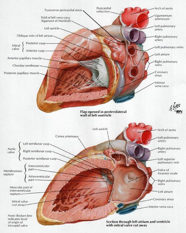
Left
ventricle is the
largest heart chamber, its wall forms larger part of diaphragmatic surface.
Internal surface containes the trabeculi carneae, which attach anterior
papillary muscle and posterior papillary muscle. The tops of these
muscles by means of cordae tendineae hold the cusps of mitral valve.
Left atrioventricular ostium
closes by left atrioventricular (bicuspidal) valve [valve mitralis],
which consists of anterior cusp and posterior cusp edges of which
attach to chordae tendineae. From left ventricle aorta starts. Aortic
ostium closes by aortic valve, which consists of right,
left and posterior semilunar valvulae, which have on superior margin the nodules
of semilunar valvulae. Between each semilunar valvula and aorta walls are
situated aortic sinuses.

Base of ventricles exposed by removal of the atria.
Size.—The heart, in the adult, measures
about
Component Parts.—As has already been
stated, the heart is subdivided by septa into right and left halves, and a
constriction subdivides each half of the organ into two cavities, the upper
cavity being called the atrium, the lower the ventricle. The
heart therefore consists of four chambers, right and left atria, and right and
left ventricles.
The division of the heart into four cavities is indicated on its surface
by grooves. The atria are separated from the ventricles by the coronary
sulcus (auriculoventricular groove); this contains the trunks of the
nutrient vessels of the heart, and is deficient in front, where it is crossed
by the root of the pulmonary artery. The interatrial groove, separating
the two atria, is scarcely marked on the posterior surface, while anteriorly it
is hidden by the pulmonary artery and aorta. The ventricles are separated by
two grooves, one of which, the anterior longitudinal sulcus, is situated
on the sternocostal surface of the heart, close to its left margin, the other posterior
longitudinal sulcus, on the diaphragmatic surface near the right margin;
these grooves extend from the base of the ventricular portion to a notch, the incisura
apicis cordis, on the acute margin of the heart just to the right of the
apex.

The base (basis cordis), directed upward, backward, and to
the right, is separated from the fifth, sixth, seventh, and eighth thoracic
vertebræ by the esophagus, aorta, and thoracic duct. It is formed mainly
by the left atrium, and, to a small extent, by the back part of the right
atrium. Somewhat quadrilateral in form, it is in relation above with the
bifurcation of the pulmonary artery, and is bounded below by the posterior part
of the coronary sulcus, containing the coronary sinus. On the right it is
limited by the sulcus terminalis of the right atrium, and on the left by the
ligament of the left vena cava and the oblique vein of the left atrium. The
four pulmonary veins, two on either side, open into the left atrium, while the
superior vena cava opens into the upper, and the anterior vena cava into the
lower, part of the right atrium.
The Apex (apex cordis).—The
apex is directed downward, forward, and to the left, and is overlapped by the
left lung and pleura: it lies behind the fifth left intercostal space, 8 to
The sternocostal surface is directed forward, upward, and to the
left. Its lower part is convex, formed chiefly by the right ventricle, and
traversed near its left margin by the anterior longitudinal sulcus. Its upper
part is separated from the lower by the coronary sulcus, and is formed by the
atria; it presents a deep concavity, occupied by the ascending aorta
and the pulmonary artery.
The diaphragmatic surface directed downward and slightly
backward, is formed by the ventricles, and rests upon the central tendon and a
small part of the left muscular portion of the diaphragm. It is separated from
the base by the posterior part of the coronary sulcus, and is traversed
obliquely by the posterior longitudinal sulcus.
The right margin of the heart is long, and is formed by the right
atrium above and the right ventricle below. The atrial portion is rounded and
almost vertical; it is situated behind the third, fourth, and fifth right
costal cartilages about
The left or obtuse margin is shorter, full, and rounded:
it is formed mainly by the left ventricle, but to a slight extent, above, by
the left atrium. It extends from a point in the second left intercostal space,
about
Right Atrium (atrium dextrum; right auricle).—The right atrium is larger than the left, but its walls are somewhat
thinner, measuring about
Sinus Venarum (sinus venosus).—The
sinus venarum is the large quadrangular cavity placed between the two
venæ cavæ. Its walls, which are extremely thin, are connected below
with the right ventricle, and medially with the left atrium, but are free in
the rest of their extent.
Auricula (auricula dextra; right auricular appendix).—The auricula is a small conical muscular pouch, the margins of which
present a dentated edge. It projects from the upper and front part of the sinus
forward and toward the left side, overlapping the root of the aorta.

Sternocostal
surface of heart.
The separation of the auricula from the sinus venarum is indicated
externally by a groove, the terminal sulcus, which extends from the front
of the superior vena cava to the front of the inferior vena cava, and
represents the line of union of the sinus venosus of the embryo with the
primitive atrium. On the inner wall of the atrium the separation is marked by a
vertical, smooth, muscular ridge, the terminal crest. Behind the crest
the internal surface of the atrium is smooth, while in front of it the muscular
fibers of the wall are raised into parallel ridges resembling the teeth of a
comb, and hence named the musculi pectinati.
Its interior presents the following parts for examination:
Openings »
Superior vena cava.
Inferior vena cava.
Coronary sinus.
Valves »
Valve of the inferior vena cava.
Foramina venarum minimarum.
Valve of the coronary sinus.
Atrioventricular.
Fossa ovalis.
Limbus fossæ ovalis.
Intervenous tubercle.
Musculi pectinati.
Crista terminalis.
The superior vena cava returns the blood from the upper half of
the body, and opens into the upper and back part of the atrium, the direction
of its orifice being downward and forward. Its opening has no valve.
The inferior vena cava, larger than the superior, returns the
blood from the lower half of the body, and opens into the lowest part of the
atrium, near the atrial septum, its orifice being directed upward and backward,
and guarded by a rudimentary valve, the valve of the inferior vena cava
(Eustachian valve). The blood entering the atrium through the superior
vena cava is directed downward and forward, i.e., toward the
atrioventricular orifice, while that entering through the inferior vena cava is
directed upward and backward, toward the atrial septum. This is the normal
direction of the two currents in fetal life.
The coronary sinus opens into the atrium, between the orifice of
the inferior vena cava and the atrioventricular opening. It returns blood from
the substance of the heart and is protected by a semicircular valve, the valve
of the coronary sinus (valve of Thebesius).

Interior
of right side of heart.
The foramina venarum minimarum (foramina Thebesii) are the
orifices of minute veins (venœ cordis minimœ), which return blood
directly from the muscular substance of the heart.
The atrioventricular opening (tricuspid orifice) is the
large oval aperture of communication between the atrium and the ventricle; it
will be described with the right ventricle.
The valve of the inferior vena cava (valvula venœ
cavœ inferioris [Eustachii]; Eustachian valve) is
situated in front of the orifice of the inferior vena cava. It is semilunar in
form, its convex margin being attached to the anterior margin of the orifice;
its concave margin, which is free, ends in two cornua, of which the left is
continuous with the anterior edge of the limbus fossæ ovalis while the
right is lost on the wall of the atrium. The valve is formed by a duplicature
of the lining membrane of the atrium, containing a few muscular fibers. In
the fetus this valve is of large size, and serves to direct the blood from
the inferior vena cava, through the foramen ovale, into the left atrium. In
the adult it occasionally persists, and may assist in preventing the reflux
of blood into the inferior vena cava; more commonly it is small, and may
present a cribriform or filamentous appearance; sometimes it is altogether
wanting.
The valve of the coronary sinus (valvula sinus coronarii [Thebesii];
Thebesian valve) is a semicircular fold of the lining membrane of the
atrium, at the orifice of the coronary sinus. It prevents the regurgitation of
blood into the sinus during the contraction of the atrium. This valve may be
double or it may be cribriform.
The fossa ovalis is an oval depression on the septal wall of the
atrium, and corresponds to the situation of the foramen ovale in the fetus. It
is situated at the lower part of the septum, above and to the left of the
orifice of the inferior vena cava.
The limbus fossæ ovalis (annulus ovalis) is the
prominent oval margin of the fossa ovalis. It is most distinct above and at the
sides of the fossa; below, it is deficient. A small slit-like valvular opening
is occasionally found, at the upper margin of the fossa, leading upward beneath
the limbus, into the left atrium; it is the remains of the fetal aperture
between the two atria

The intervenous tubercle (tuberculum intervenosum; tubercle of
Lower) is a small projection on the posterior wall of the atrium, above the
fossa ovalis. It is distinct in the hearts of quadrupeds, but in man is
scarcely visible. It was supposed by Lower to direct the blood from the
superior vena cava toward the atrioventricular opening.
Right Ventricle (ventriculus dexter).—The
right ventricle is triangular in form, and extends from the right atrium to
near the apex of the heart. Its anterosuperior surface is rounded and convex,
and forms the larger part of the sternocostal surface of the heart. Its under
surface is flattened, rests upon the diaphragm, and forms a small part of the
diaphragmatic surface of the heart. Its posterior wall is formed by the
ventricular septum, which bulges into the right ventricle, so that a transverse
section of the cavity presents a semilunar outline. Its upper and left angle
forms a conical pouch, the conus arteriosus, from which the pulmonary
artery arises. A tendinous band, which may be named the tendon of the conus
arteriosus, extends upward from the right atrioventricular fibrous ring and
connects the posterior surface of the conus arteriosus to the aorta. The wall
of the right ventricle is thinner than that of the left, the proportion between
them being as 1 to 3; it is thickest at the base, and gradually becomes thinner
toward the apex. The cavity equals in size that of the left ventricle, and is
capable of containing about 85 c.c.
Its interior presents the following parts for examination:
Openings »
Right atrioventricular.
Valves »
Tricuspid.
Pulmonary artery.
Pulmonary.
Trabeculæ carneæ
Chordæ tendineæ
The right atrioventricular orifice is the large oval aperture of
communication between the right atrium and ventricle. Situated at the base of
the ventricle, it measures about
The opening of the pulmonary artery is circular in form, and
situated at the summit of the conus arteriosus, close to the ventricular
septum. It is placed above and to the left of the atrioventricular opening, and
is guarded by the pulmonary semilunar valves.
The tricuspid valve (valvula
tricuspidalis) consists of three somewhat triangular cusps or segments. The
largest cusp is interposed between the atrioventricular orifice and the conus
arteriosus and is termed the anterior or infundibular cusp. A
second, the posterior or marginal cusp, is in relation to the
right margin of the ventricle, and a third, the medial or septal
cusp, to the ventricular septum. They are formed by duplicatures of the
lining membrane of the heart, strengthened by intervening layers of fibrous
tissue: their central parts are thick and strong, their marginal portions thin
and translucent, and in the angles between the latter small intermediate
segments are sometimes seen. Their bases are attached to a fibrous ring
surrounding the atrioventricular orifice and are also joined to each other so
as to form a continuous annular membrane, while their apices project into the
ventricular cavity. Their atrial surfaces, directed toward the blood current
from the atrium, are smooth; their ventricular surfaces, directed toward the
wall of the ventricle, are rough and irregular, and, together with the apices
and margins of the cusps, give attachment to a number of delicate tendinous
cords, the chordæ tendineæ.


Heart
seen from above.
The trabeculæ carneæ (columnœ carneœ)
are rounded or irregular muscular columns which project from the whole of the
inner surface of the ventricle, with the exception of the conus arteriosus.
They are of three kinds: some are attached along their entire length on one
side and merely form prominent ridges, others are fixed at their extremities
but free in the middle, while a third set (musculi papillares) are
continuous by their bases with the wall of the ventricle, while their apices
give origin to the chordæ tendineæ which pass to be attached to the
segments of the tricuspid valve. There are two papillary muscles, anterior and
posterior: of these, the anterior is the larger, and its chordæ
tendineæ are connected with the anterior and posterior cusps of the
valve: the posterior papillary muscle sometimes consists of two or three parts;
its chordæ tendineæ are connected with the posterior and medial
cusps. In addition to these, some chordæ tendineæ spring directly
from the ventricular septum, or from small papillary eminences on it, and pass
to the anterior and medial cusps. A muscular band, well-marked in sheep and
some other animals, frequently extends from the base of the anterior papillary
muscle to the ventricular septum. From its attachments it may assist in
preventing overdistension of the ventricle, and so has been named the moderator
band.
The pulmonary semilunar valves are three in number, two in front
and one behind, formed by duplicatures of the lining membrane, strengthened by
fibrous tissue. They are attached, by their convex margins, to the wall of the
artery, at its junction with the ventricle, their free borders being directed
upward into the lumen of the vessel. The free and attached margins of each are
strengthened by tendinous fibers, and the former presents, at its middle, a
thickened nodule (corpus Arantii). From this nodule tendinous fibers
radiate through the segment to its attached margin, but are absent from two
narrow crescentic portions, the lunulæ, placed one on either side
of the nodule immediately adjoining the free margin. Between the semilunar
valves and the wall of the pulmonary artery are three pouches or sinuses
(sinuses of Valsalva).

Left Atrium (atrium sinistum; left auricle).—The left atrium is rather smaller than the right, but its walls are
thicker, measuring about
The principal cavity is cuboidal in form, and concealed, in
front, by the pulmonary artery and aorta; in front and to the right it is
separated from the right atrium by the atrial septum; opening into it on either
side are the two pulmonary veins.
Auricula (auricula sinistra; left auricular appendix).—The auricula is somewhat constricted at its junction with the principal
cavity; it is longer, narrower, and more curved than that of the right side,
and its margins are more deeply indented. It is directed forward and toward the
right and overlaps the root of the pulmonary artery.
The interior of the left atrium presents the following parts for
examination:
Openings of the four pulmonary veins.
Left atrioventricular opening.
Musculi pectinati.
The pulmonary veins, four in number, open into the upper part of
the posterior surface of the left atrium—two on either side of its middle line:
they are not provided with valves. The two left veins frequently end by a
common opening.
The left atrioventricular opening is the aperture between the
left atrium and ventricle, and is rather smaller than the corresponding opening
on the right side.
The musculi pectinati, fewer and smaller than in the right
auricula, are confined to the inner surface of the auricula.
On the atrial septum may be seen a lunated impression, bounded below by
a crescentic ridge, the concavity of which is turned upward. The depression is
just above the fossa ovalis of the right atrium.
Left Ventricle (ventriculus sinister).—The left ventricle is longer and more conical in shape than the right,
and on transverse section its concavity presents an oval or nearly circular
outline. It forms a small part of the sternocostal surface and a considerable
part of the diaphragmatic surface of the heart; it also forms the apex of the
heart. Its walls are about three times as thick as those of the right
ventricle.
Its interior presents the following parts for examination:
Openings »
Left atrioventricular.
Valves »
Bicuspid or Mitral.
Aortic.
Aortic.
Trabeculæ carneæ.
Chordæ tendineæ
The left atrioventricular opening (mitral orifice) is
placed below and to the left of the aortic orifice. It is a little smaller than
the corresponding aperture of the opposite side, admitting only two fingers. It
is surrounded by a dense fibrous ring, covered by the lining membrane of the
heart, and is guarded by the bicuspid or mitral valve.

Interior
of left side of heart.

Aorta
laid open to show the semilunar valves.
The aortic opening is a circular aperture, in front and to the
right of the atrioventricular, from which it is separated by the anterior cusp
of the bicuspid valve. Its orifice is guarded by the aortic semilunar
valves. The portion of the ventricle immediately below the aortic orifice
is termed the aortic vestibule, and possesses fibrous instead of
muscular walls.
The bicuspid or mitral valve (valvula bicuspidalis
[metralis]) is attached to the circumference of the left
atrioventricular orifice in the same way that the tricuspid valve is on the
opposite side. It consists of two triangular cusps, formed by duplicatures of
the lining membrane, strengthened by fibrous tissue, and containing a few
muscular fibers. The cusps are of unequal size, and are larger, thicker, and
stronger than those of the tricuspid valve. The larger cusp is placed in front
and to the right between the atrioventricular and aortic orifices, and is known
as the anterior or aortic cusp; the smaller or posterior cusp
is placed behind and to the left of the opening. Two smaller cusps are usually
found at the angles of junction of the larger. The cusps of the bicuspid valve
are furnished with chordæ tendineæ, which are attached in a manner
similar to those on the right side; they are, however, thicker, stronger, and
less numerous.
The aortic semilunar valves are three in number, and surround the
orifice of the aorta; two are anterior (right and left) and one posterior. They
are similar in structure, and in their mode of attachment, to the pulmonary
semilunar valves, but are larger, thicker, and stronger; the lunulæ are
more distinct, and the noduli or corpora Arantii thicker and more prominent.
Opposite the valves the aorta presents slight dilatations, the aortic
sinuses (sinuses of Valsalva), which are larger than those at the
origin of the pulmonary artery.
The trabeculæ carneæ are of three kinds, like those
upon the right side, but they are more numerous, and present a dense
interlacement, especially at the apex, and upon the posterior wall of the
ventricle. The musculi papillares are two in number, one being connected
to the anterior, the other to the posterior wall; they are of large size, and
end in rounded extremities from which the chordæ tendineæ arise.
The chordæ tendineæ from each papillary muscle are connected to
both cusps of the bicuspid valve.

Section
of the heart showing the ventricular septum.
Ventricular Septum (septum ventriculorum; interventricular septum)
The ventricular septum is directed obliquely backward
and to the right, and is curved with the convexity toward the right ventricle:
its margins correspond with the anterior and posterior longitudinal sulci. The
greater portion of it is thick and muscular and constitutes the muscular
ventricular septum, but its upper and posterior part, which separates the
aortic vestibule from the lower part of the right atrium and upper part of the
right ventricle, is thin and fibrous, and is termed the membranous
ventricular septum. An abnormal communication may exist between the
ventricles at this part owing to defective development of the membranous
septum.


Purkinje’s fibers
from the sheep’s heart. A.
In longitudinal section. B. In transverse section.

The fibers of the ventricles are arranged
in a complex manner, and various accounts have been given of their course and
connections; the following description is based on the work of McCallum. They consist of superficial and deep layers,
all of which, with the exception of two, are inserted into the papillary
muscles of the ventricles. The superficial layers consist of the
following: (a) Fibers which spring from the tendon of the conus
arteriosus and sweep downward and toward the left across the anterior
longitudinal sulcus and around the apex of the heart, where they pass upward
and inward to terminate in the papillary muscles of the left ventricle; those
arising from the upper half of the tendon of the conus arteriosus pass to the
anterior papillary muscle, those from the lower half to the posterior papillary
muscle and the papillary muscles of the septum. (b) Fibers which arise
from the right atrioventricular ring and run diagonally across the
diaphragmatic surface of the right ventricle and around its right border on to
its costosternal surface, where they dip beneath the fibers just described,
and, crossing the anterior longitudinal sulcus, wind around the apex of the
heart and end in the posterior papillary muscle of the left ventricle. (c)
Fibers which spring from the left atrioventricular ring, and, crossing the
posterior longitudinal sulcus, pass successively into the right ventricle and
end in its papillary muscles. The deep layers are three in number; they
arise in the papillary muscles of one ventricle and, curving in an S-shaped manner,
turn in at the longitudinal sulcus and end in the papillary muscles of the
other ventricle. The layer which is most superficial in the right ventricle
lies next the lumen of the left, and vice versa. Those of the first
layer almost encircle the right ventricle and, crossing in the septum to the
left, unite with the superficial fibers from the right atrioventricular ring to
form the posterior papillary muscle. Those of the second layer have a less
extensive course in the wall of the right ventricle, and a correspondingly
greater course in the left, where they join with the superficial fibers from
the anterior half of the tendon of the conus arteriosus to form the papillary
muscles of the septum. Those of the third layer pass almost entirely around the
left ventricle and unite with the superficial fibers from the lower half of the
tendon of the conus arteriosus to form the anterior papillary muscle. Besides
the layers just described there are two bands which do not end in papillary
muscles. One springs from the right atrioventricular ring and crosses in the
atrioventricular septum; it then encircles the deep layers of the left
ventricle and ends in the left atrioventricular ring. The second band is
apparently confined to the left ventricle; it is attached to the left
atrioventricular ring, and encircles the portion of the ventricle adjacent to
the aortic orifice.
Dr. A. Morison has shown that in the sheep and pig the atrioventricular bundle “is a
great avenue for the transmission of nerves from the auricular to the
ventricular heart; large and numerous nerve trunks entering the bundle and
coursing with it.” From these, branches pass off and form plexuses around
groups of Purkinje cells, and from these plexuses fine fibrils go to innervate
individual cells.

The lymphatics end in the thoracic
and right lymphatic ducts.


-
lesser cardiac vein [vena cordis parva], which passes in right part
of coronal sulcus;
-
middle cardiac vein [vena
cordis media] passes
in posterior interventricular sulcus;
-
posterior vein of left ventricle;
-
oblique vein of left atrium.
There are venae minimae
(Tebezia) and anterior venae, positioned in myocardium of right atrium.


